Saturday, September 28, 2013

Ancient Parasites of Puppies in Egypt

Researchers showing the infested ear of the mummified dog.
A really great article came out in the International Journal of Paleopathology last month.  It was entitled, "The dog mummy, the ticks and the louse fly: Archaeological report of severe ectoparasitosis in Ancient Egypt".  In this paper, a site in El Deir, Egypt was excavated between 2010 and 2011.  There were hundreds of mummified dogs found at the site. One of the dog mummies was unique from the others...it had ectoparasites!
This dog represents the first real evidence of canine ectoparasitism in Ancient Egypt. Researchers found a great load of ticks and a louse fly still attached to the ears and coat of the mummified pup.  These parasites may have been vectors for a variety of diseases that could have lead to the early passing of the young dog.


Mummified dog's ear infested with ticks.

People have suspected that such diseases existed in antiquity based on the writings of early Greek and Latin scholars.  Aristotle called do parasites kunorhaistes, a.k.a "dog destroyer".  Homer described Ulysses' dog as being infested by the same.  Pliny the Elder also described ticks that burdened dogs of his day saying:

"There is an animal...that always lives with its head fixed in the blood of a host, and consequently goes on swelling, as it is the only animal that has no vent for its food; with gorging to excess it bursts, so dying of its very nutriment.  This creature...occurs frequently in oxen and occasionally in dogs in which all creatures breed."
 
The oldest record of ticks exists in a piece of art dating back to Ancient Egypt.  This art depicts a "hyena-like animal" with ticks in its ear and dates to the 15th century B. C.  The piece is currently housed in the Metropolitain Museum of Art in New York.  This new paper represents the first time that anyone has actually found hard evidence that such ectoparasites did, in fact, exist. 

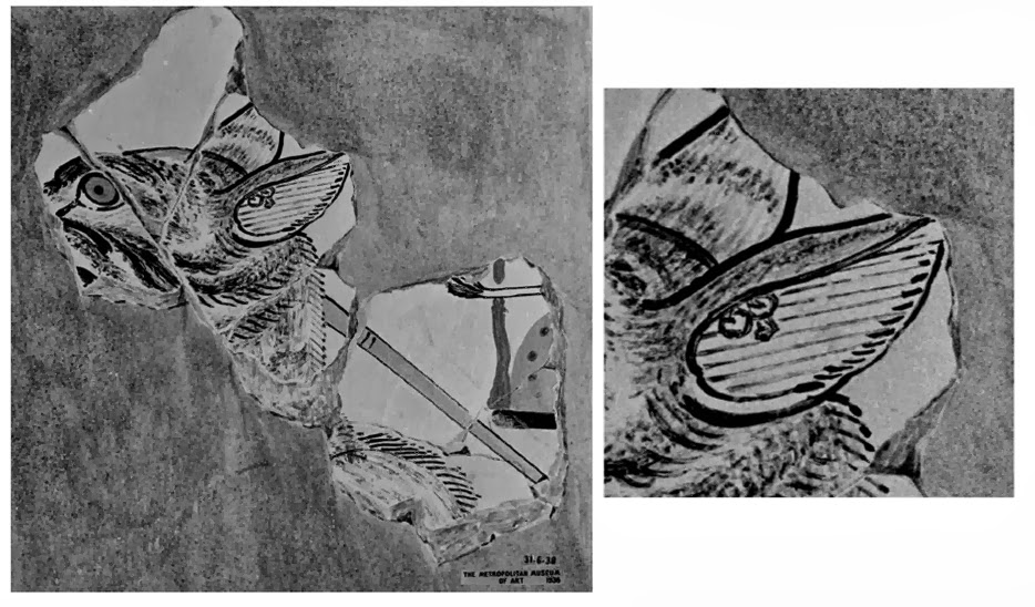
Piece from the Tomb of Intef, New Kingdom, Thebes, Upper Egypt.
Okay, parasite details!  Parasite details!  The ectoparasites found fell into two types.  The first was the brown dog tick, Rhipicephalus sanguineus, a type of hard tick found commonly all over the world, but especially in warm climates.  The second type was the louse fly, Hippobosca longipennis, a type of hippoboscid fly native to Africa, Asia, and the Middle East. 


There were 61 individual specimens of the hard tick recovered from the dog mummy with 38% of them pulled out of the inner ear alone.  This indicates that the 4-5 month old puppy may have suffered from problems like anemia or tick-borne pathogens.  Only a single louse fly was found on the coat of the animal.

The researchers also found the puparia of flies belonging in the families of Calliphoridae and Sarcophagidae.  The presence of these puparia could have been a source of myiasis (the infection of a live mammal with the larvae  of these flies).  However, the presence of these flies may also have invaded the dog mummies post-mortem. (Which I, personally, feel is more likely.)

Moral of the Story
It is pretty exciting to find solid proof of canine parasitism dating back to Ancient Egypt.  That being said, I have to wonder whether or not a parasitologist was a part of this research.  I have a few questions that come to mind about this amazing ectoparasite discovery.  First and foremost, why the hell were the ticks still attached to the dog?  Usually, when a tick's host dies, the ticks...to the best of my limited knowledge...abandon ship and go off questing for another host with their little jointed appendages reaching out for love.  Yet, here we find amazingly well-preserved ticks that, quite literally, had a death grip on the ears of these dogs.  WHY?  The authors proposed that the ticks were still there because their hypostomes have been known to sort of get stuck from time to time and stay attached to their hosts.  (They did not cite this assertion, so I'm not sure where that has been reported.)  Sure, the ticks might not pop off immediately, but I doubt they would hang on to the point that they would die and become mummified along with their host.  I'm thinking there had to be something else going on here!  The authors also proposed that perhaps the parasites were vectors for diseases that led to the early demise of the dog.  Though completely plausible, I'm not sure that I totally buy into this idea.  

Parasites and puparia collected
from dog mummies.
Another question that came to mind for me was the authors' theory that perhaps the puparia belonged to flies that had invaded their hosts' tissues while the hosts were still alive. I'm not really buying that one either.  I believe that it is far more likely that these puparia belonged to flies that laid eggs/larvae in their hosts post-mortem.  After hatching and molting, they would have pupated prior to some abrupt happenstance that preserved them before they could complete their life cycle.  (Although many of the puparia were fragments rather than whole puparia, indicating that many of the flies may have completed their life cycle without hindrance.)  It seems to me that perhaps the dog died in some way that killed the ticks and louse fly as quickly as it killed the dogs (perhaps a strong poison of some sort?) or possibly the dog became paralyzed (via ticks or from whatever caused the three vertebral dislocations I didn't mention earlier) and the owners entombed the dog thinking it was dead.   In either case, perhaps the dog's body was left exposed long enough for flesh flies and blow flies to invade its corpse.  Afterwards, the dog was placed in what I'm speculating may have been an anaerobic environment that could have halted the lives of the flies just after reaching their pupal stages.  This would be one explanation for the presence of these flies and arachnids found in these mummies.

My biological curiosities aside, it is very important that we can definitively say that the ectoparasitism of domesticated dogs dates back to the days of Ancient Egypt.  This is an incredible find in that it tells us that the host-parasite interactions found in modern canines are not all that different from those found thousands of years ago.  It is also interesting to note that the morphology of the brown dog tick hasn't changed much in all that time...I suppose if it ain't broke... This research will be helpful for those who are especially interested in the evolution of canine parasitism. Hooray for paleoparasitology!!!

Saturday, September 21, 2013

Creepy Crawlers for Crohn's: Coming to a Scientific Journal Near You!

I've mentioned helminthic therapy and the lost friends theory on this blog before. (I'm not lying! Click here or here if you don't believe me!) As I was browsing the interwebs I ran across a short article about the use of pig whipworms (Trichuris suis) in treating Crohn's disease. Of course, I smiled...I love the lost friends theory and I've read that helminthic therapy has remission rates in the 90s for people with Crohn's and ulcerative colitis.  The article was pretty standard...it works pretty well...there's no autoinfeciton...the biggest hurdle with patients is the "yuck" factor...but one of the last paragraphs of the article really caught my attention:

"We’ll know more about the whipworm’s effectiveness when results of a phase 2 clinical trial involving 250 patients with Crohn’s disease is published in the fourth quarter of this year. Coronado also is testing its parasite therapy in patients with multiple sclerosis, ulcerative colitis and psoriasis, and expects to begin a type 1 diabetes trial soon." --This Article

Just a Petri dish of little whipworms.
I almost jumped off of my couch and broke my laptop that I can't really afford to replace. (Good thing almost doesn't count!....except of course in horseshoes and hand grenades.)  They have 250 patients with Crohn's in phase 2 of clinical trials with helminthic therapy?!?!?! HOW COOL IS THAT?!?!?!  I'm super stoked to see the results of the study!  With good, solid clinical trials nearing completion, this means that helminthic therapy will gain notoriety among doctors as an effective treatment option!  (Assuming it works...which, by all accounts I've read it does....the question is whether or not it is more effective that conventional drugs...which, according to everything I've read on patient forums, all signs point to yes...and *bonus points* it's far cheaper than pharmaceuticals.) I can't wait for this paper to hit scientific journals so I can be blown away all over again.  This really is revolutionary...it's an exciting time to be a parasitologist!  Finally, some good press for the things we parasitophiliacs love and adore! I'm probably getting a little ahead of myself...let us wait and see what the results tell us!

Also, I've read about helminthic therapy for multiple sclerosis and of course for ulcerative colitis, but the psoriasis thing is new!  I have also read about the use of helminthic therapy for food allergies...it's pretty amazing how helpful these little guys can be.  The last sentence really, really blew me away..."expects to begin a type 1 diabetes trial soon"???  So we can add diabetes to the list of things that might be treatable by purposely infecting oneself with parasites? DIA-FREAKIN-BETES?!?!?! WOW!!!!  An exciting time to live indeed!

Diagram showing common skin bacteria...just the skin...
While I'm here, here's a little note on the "yuck" factor: If you are a person suffering from Crohn's disease, ulcerative colitis, or good lord multiple sclerosis, are you really going to let your mind freak you out over a treatment that is cheaper than drugs, has high remission rates, and causes fewer severe side effects?  With the pain you are living in and the debt you are accruing, what is keeping you from giving this type of therapy a try?  Is the thought of something living inside of you really that disturbing?  Do you realize that you already have things...LOTS of things...living inside of your body RIGHT THIS MOMENT???  Your body depends on an array of gut microbes in order to even function.  Millions of bacteria are colonizing your body and helping you to do things your body could never accomplish on its own.  You've also likely played host to hundreds of things in your lifetime...most of which you developed antibodies to in order to help yourself fight off such invaders the next time they come around. You've also (hopefully) had things introduced into your body to help you build these antibodies in order to protect you from getting full-on diseases like measles, diptheria, and polio.  Your body is a walking skin-bag of microorganisms zipping through it to keep the well-oiled machine of you going.  If some part of your body is having maintenance issues, and costly prescriptions and surgeries aren't helping, why not call in some wormy-friends to kick-start your immune system?  After all, what have you got to lose? Get over the idea of "something living inside of me" and get yourself feeling better!

Moral of the Story
Be on the lookout for the new paper about the effectiveness of helminthic therapy in treating Crohn's disease!  Coming to a peer-reviewed journal near YOU in the fourth quarter of this year! :)


Sunday, September 15, 2013

Toxo-Tainted Meats

Recalling a recent conversation in which an undergrad accused me of "stealing [her] parasite" (she was a touch inebriated, so we will cut her some slack on this eyebrow-raising comment), I decided to do a post on one of my favorite parasites.  I've blogged about this parasite before (here and here), so today I'll be discussing a bit about the prevalence of the parasite in animals often destined for human consumption.  I'm speaking of course, about Toxoplasma gondii.

To get ourselves oriented appropriately, let's start by looking at the range of hosts known to harbor T. gondii. Though the typical life cycle involves a hungry cat and a brainwashed mouse, T. gondii actually has quite a broad range of hosts.  It has been known to infect a wide variety of rodents as well as birds, bats, deer, bears, sea otters, and rabbits.  The prevalence of toxoplasmosis in those animals ranges between 50% and 70% in some populations.  Domesticated dogs can get toxoplasmosis, but domesticated cats are much more likely to become infected.  Livestock such as cattle, goats, pigs, and sheep are also target hosts for this parasite.  Infection rates in livestock may have prevalence rates of 50% or higher.  Humans can also become infected (and they do!).  An estimated 30% of the world population is infected with T. gondii, while the same parasite infects about 22% of the U.S. population.

Not the best resolution, but you can tell from the pictures that people may become infected in a variety of ways.

Okay, so now let's focus on the stuff we might be eating.  Like I mentioned a moment ago, this parasite can be found in many forms of livestock. I recently read an article about the prevalence of this parasite in chickens as well.  That article was comparing prevalence rates of free-range chickens with those of cage-raised chickens.  You would think that cage-raised chickens would have a higher prevalence since they live in closer contact with other chickens.  While this is certainly true for a number of diseases that plague the poultry industry, this is not the case with toxoplasmosis.  Instead, free-ranged chickens had twice the prevalence of cage-raised chickens.  The theory is that free-range chickens are more likely to come into contact with wild hosts like field mice, rabbits, etc.

There are also several papers documenting the occurrence of toxoplasmosis in free-range beef, mutton, and especially in pork.  Unlike with the chicken article, most of these papers were not comparing free-range animals with their caged counterparts.  Instead, the authors of these papers were simply looking at prevalence.  Most of the papers were reporting prevalence rates well over 50%.  However, the question remains...how much of that tainted meat actually makes it to market?

A survey of supermarkets in Taipei showed the prevalence of Toxoplasma was 10% in pork and other pig products, 4% in mutton, 6% in chicken and chicken products, and 5% in beef.  This survey just came out this year.  Another study conducted in 2005 concluded that U.S. meat retailers also had low prevalences of viable T. gondii oocysts. (The majority of tainted meats were of pork origin.)

This graphic came from this article.  The graphic shows the relative importance
of consumed animals in the transmission of toxoplasmosis to humans.

While these numbers aren't staggering, they are still present.  Consumers, particularly pregnant or otherwise immunocompromised consumers, should be aware of the fact that they can contract this parasite even from free-range meat sources.  Luckily, you can avoid getting the parasite even if you do purchase tainted meat by simply ensuring that your meats are cooked to an internal temperature of 150.8 degrees F (66 degrees C) in order to kill Toxoplasma.

Moral of the Story
While there are lots of benefits to consuming free-range meats, this does not mean that you no longer have to worry about parasites in your food.  Always properly cook your burgers, gizzards, and pulled pork...especially if you are prego or if you have AIDS.  Just another reason to try cultured beef when it finally hits the shelves here in 10 years or so...petri-dished meat can't contract toxoplasmosis unless some lab tech (who would shortly cease to be employed) purposefully infected a batch.  Even then, the unemployed, idiotic lab tech wouldn't get anyone sick as long as consumers cooked the meat long enough!


Monday, September 9, 2013

Parasitic Jellyfish?!?!?! Meet Polypodium hydriforme!

Polypodium hydriforme...a parasitic cnidarian!
I had the great fortune to attend a great regional parasite conference this past weekend.  The RockyParasitophile in bold, white lettering above the name of the conference, with the hosting field station's logo on the back. The shirts themselves were black and over 2/3rds of the order sold in the three days of the conference!)  At one point, I wound up sitting at a table with a fellow PhD student and two well-known parasitologists who teach at major universities.  During what was an exciting conversation (in which I was mostly trying to listen instead of speaking), one of the professors mentioned a parasitic cnidarian! I was so stunned I didn't pipe up before the conversation was on to other things.  (I was also a bit slow since this was around 2am.) Luckily, I was able to catch one of the professors the next day and ask for the scientific name of the parasite.  So, without further ado, meet the most uniquely awesome jellyfish of all time...Polypodium hydriforme!
Mountain Conference of Parasitologists is a great way to learn more about parasites and to meet fellow parasitophiles.  (Actually, I was able to help design a RMCP t-shirt for this year's conference...it had the word

Taxonomy
Diagram of a cnidocyst.
Unlike every other parasite I've ever blogged about here on Parasitophilia, this parasite belongs to phylum Cnidaria.  Cnidaria is the phylum in which you find animals that are diploblastic (unlike the other things we've talked about, which are all triploblastic).  This group includes animals like jellyfish, sea anemones, tiny freshwater guys like Hydra and Obelia.  All members have tentacles with stinging cells called cnidocytes.  The cnidocytes are equipped with nematocysts (stinging capsules) that are fired at prey items when the cnidarians are ready to feed.  P. hydriforme has these structures, just like other cnidarians.  The parasite also belongs to the class Polypodiozoa and the family Polypodiidae.  This family only contains one species (that's right, this little guy has a whole family to itself)!  Obviously, it is the only member of this genus as well.

Recent molecular studies have brought the conventional taxonomic categorization for this organism into question.  There is some evidence that this parasite may be more closely related to myxozoans than to cnidarians.  However, the presence of cnidocytes still makes this organism a cnidarian in my classically-trained, organismally-based, little biologist eyes.


Normal, black eggs among infected eggs. The arrows show
abnormal eggs and the circle denotes a mature stolon.
Life Cycle
The life cycle of this parasite begins with the emergence of the parasite from the eggs of its host organism, a type of sturgeon or paddlefish.  The parasite emerges in a life stage known as a "stolon" from the eggs in a fresh water ecosystem.  The stolon fragments into a bunch of tiny little medusa-like stages (when you think "medusa" stages....think of the morphology of what you normally see when you look at jellyfish in the zoo).  These little medusoid forms go on to multiply by splitting in half and then growing sexual organs. Eventually, the parasites release their gametes, which mate in the water to form an embryo.  The embryo develops into a planuliform larva, which then infects the bodies of the appropriate host fish.  Within the bodies, the parasite infects the oocytes, where it lives the majority of its life. The larvae develop into an inside-out stolon and waits until the fish is ready to spawn.  Just before the fish releases its eggs, the stolon everts itself to reveal its tentacles (within the egg).  After being released, the eggs become the source of food for the parasite for a time before the stolon emerges from the eggs. (This is the best interpretation of the life cycle that I could understand, if you see something that isn't accurate, please let me know!)

A mature stolon with everted tentacles.
Economic Importance
One of the things that makes this parasite especially important (beside the fact that it's really cool!), is that it infects the eggs of fish that have culinary significance.  One of the biggest problems is that sturgeons contract these parasites.  Sturgeons, for those of you who don't know, produce eggs that many people eat as a delicacy....a very expensive delicacy...it's caviar...caviar is sturgeon eggs.  These eggs are normally small and black, but when infected by P. hydriforme the eggs become enlarged and take on a gray appearance.  If this parasite gets into a farm that raises sturgeons to harvest caviar, it can wreck the farm's production levels.  Many wealthy connoisseurs would be distraught without their caviar...though personally, I'm not a fan of sturgeon eggs.  I really couldn't care less if the caviar industry died off.  However, I would be very sad if that meant the end of the sturgeon.  Sturgeons don't deserve to go extinct.  Then again, I doubt that if the market for caviar disappeared that the fish would disappear alongside it.  They might be doing just fine in the wild.  (I can't confirm that though, you'll have to ask an ichthyologist.)

Another mature, free-living stolon.

Moral of the Story 
What it all comes down to is this...we have an awesomely unique parasite that we know a little more about now!  It impacts the caviar industry, but not so heavily that it stops caviar production. I suppose having less caviar means they can charge more for it anyway (because caviar isn't expensive enough as it is).  Perhaps the parasite is good for sturgeon egg farmers needing an excuse to boost their prices?!  Or perhaps the parasite is just awesome for being a parasitic cnidarian/myxozoan (depending on your taxonomic perspective).  Either way, Polypodium hydriforme is one amazing parasite that every parasitophile should know a little something about!

Sunday, September 1, 2013

Triumphs and Tragedies: The Battle Rages with Naeglaria

I've posted about this parasite before.  It's a tiny, single-celled organism that, although rare, is extremely dangerous.  I speak of course about the notorious Naeglaria fowleri.  As global temperatures rise, so do the ambient surface temperatures in lakes and other bodies of water.  This amoeba is typically non-parasitic, living in the sediments of many lakes.  However, when temperatures rise, these amoebas undergo a morphological change into flagellated forms.  These new forms are opportunistic parasites that infect unsuspecting swimmers by riding tiny tides of water into the nasal cavities of their hosts.  From there, the parasites make their way into the brain, wrecking everything in their path. 

Infection with this parasite is estimated to be between 97% and 99% fatal.  There have only been two survivors out of the 130+ cases reported in North America since 1962.  One survivor was an American and the other was from Mexico.  This summer, we mourn the passing of yet another victim, 12-year-old Zachary Reyna from LaBelle, Florida.  Zachary was infected on August 3rd after knee-boarding with friends in a drainage ditch near his home.  He was taken to Miami Children's Hospital and put on both antibiotics and on miltefosine, a German-manufactured drug often prescribed for treating breast cancer.  By August 21st, the boy's family announced that the drugs were successfully slowing the activity of the parasites, but unfortunately Zachary had stopped showing signs of brain activity.  A few weekends ago, his family had to make the tough decision to take him off of life support.  Zachary's organs were donated to help those in need.

Despite this awful tragedy, this summer also brought a triumph in the battle against this deadly parasite.  A few states away and about a month before Zachary's infection, Kali Hardig contracted this same parasite from a water park in central Arkansas. Kali is also 12 years old and was admitted to Arkansas Children's Hospital on July 19th with a severe fever.  Kali's treatment began with doctors cooling her body in order to reduce the swelling instigated by the infection.  Kali was then treated with miltefosine, which would later be used to treat Zachary.  For the next two weeks Kali was on a ventilator.  She is now breathing on her own and is responsive, though she had not been able to speak yet as of August 14th (the date of the report I was reading).  Tests have confirmed that she is parasite free and doctors say she will survive but will need weeks of rehabilitation.  This triumph makes Kali survivor #3 since 1962.  To learn more about Kali and her story, check out the Prayers for Kali Le Ann Facebook page.

Before I sign off, let's talk a little bit about Miltefosine.  This drug was originally developed as a chemotheraputic used to fight cancerous tumors.  It achieves this by inhibiting Akt (a.k.a. Protein Kinase B), which plays an important role in glucose metabolism, transcription, apoptosis (programed cell death), cell migration, and cell proliferation.  It is easy to see how this would work for treating cancerous cells.  In recent years, the drug has been found to be an effective antiprotozoal drug.  Just like with cancerous cells, unicellular parasitic protists can't survive if Akt is inhibited within their cellular membranes.  The drug has been show to be effective against leishmaniasis, trichomoniasis, Chagas' disease (in animal studies), a variety of fungal infections (caused by Aspergillus, Candida, Cryptococcus, and Fusarium), and free-living amoeba infections (caused by Acanthamoeba, Balamuthia mandrillaris, and as you've just learned Naegleria fowleri).  A compound that is similar to miltefosine structurally (hexadecyltrimethylammonium bromide) is also showing signs of effectiveness against Plasmodium falciparum, the most severe form of malaria.

Moral of the Story
It's important that we continue to advance our medical technologies through continued research.  Every day we are getting closer to finding a cure for diseases like primary amoebic meningitis (caused by Naeglaria fowleri).  Through tragedy we are reminded that we still have a long way to go, but through triumphs we are reminded that we are at least on the right track.  With the summers becoming more and more intense, it is important that we understand how to prevent these often fatal infections just as it is vital that we invest the time, money, and energy into find a way to defeat the parasite when it does manage to infect our loved ones.