Sunday, December 29, 2013

A Fluke, a Tapeworm, and a Roundworm Walk into a Sushi Bar...

I know what you are thinking. You are thinking, "I can't read this blogpost because I LOVE sushi and she WILL NOT ruin it for me!" Don't worry, I'm a big fan of sushi too, and I certainly don't want to diminish the amazingness that is this Japanese delicacy. Let me start off by saying that most of the time, especially here in the U.S. or in countries with well-regulated sushi bars (such as is the case in Japan), you are not at risk for contracting parasites from eating sushi. I'll end this post with a brief discussion about raw fish regulations just to ease your troubled mind. That being said, let's talk about the parasites that you can get from eating raw fish that hasn't been properly processed.

There are many different species of parasites that use fish as one of their hosts. Any of these parasites has the potential to infect humans if accidentally eaten. You can pick up a variety of flukes, tapeworms, and roundworms from a variety of marine and freshwater fish. To keep this blogpost to a reasonable size, we will only look at one representative from each of these three groups. We will talk about the fluke Clonorchis sinensis, the tapeworm Diphyllobothrium latum, and the roundworm Anisakis simplex.

Because we are looking at three different worms under the theme of "can be in sushi", I won't go into the detail that I normally do. I've never blogged about C. sinensis or about A. simplex, but you can find a previous blogpost about D. latum here. (Perhaps I'll blog about the other two in later posts.)

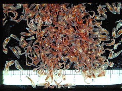
Clonorchis specimens from a patient.
These three parasites not only represent three different classes of organisms (and two different phyla if you are keeping up taxanomically), they also represent parasites found in different types of fish. C. sinensis is typically found in freshwater fish or in fish that prefer brackish waters (a mix between freshwater and marine ecosystems). This parasite is really only found in East Asia, where it utilizes a snail for its first intermediate host and a fish as its second intermediate host. The parasite has been known to be problematic in regions that import fish from East Asia in addition to popping up in local populations where it is endemic. It is also interesting to note that this parasite has been identified in mummies and coprolites from Korea. This tells us that humans have a long history of association with this particular parasite.

The fish tapeworm, D. latum, also boasts a long association with humans. Coprolites from both North America and from South America have tested positive for this parasite. Some of the earliest New World human populations were infected with fish tapeworms, which makes sense given their proximity to water sources and diets that often integrated fish. A diet that included fish is evidenced by the existence of bones and scales in macroscopic remains from coprolites as well as in artifacts constructed from fish bones. This parasite infects freshwater fish, such as trout, and can be found just about anywhere in the world. It is often diagnosed in campers/fishermen who do not properly cook their catches and in sweet little old Jewish ladies who taste test tainted gefilte fish before the dish is fully cooked.


Anisakis worms embedded in a herring.
The last of the three, A. simplex, is by far the most notorious. This roundworm is cosmopolitan in nature, like D. latum, but prefers for its hosts to be marine fish as opposed to freshwater fish. It is most often associated with mackerel and herring, which has earned it the common name of "herring worm". Apparently it can infect many other marine fish and even things like squid. It has been contracted from dishes around the world including sushi/sashimi, cod livers, fermented herring, and ceviche. Though a person can experience mild to moderate abdominal pain after contracting one of the other two parasites mentioned here, a person contracting A. simplex will experience much more violent abdominal pains. These pains are sudden and severe by comparison because these worms actually die when they fail at their attempts to burrow into your intestines.  This often instigates an IgE-mediated immune response (i.e. an allergic reaction, sometimes even anaphylaxis), making this parasite by far the most dangerous of the three discussed in this blogpost.

Ceviche
There are a great variety of symptoms to look out for if you think you may have picked up one of these parasites. Because they affect the digestive system, you may experience things like abdominal pain, vomiting, nausea, loss of appetite, and diarrhea. The first of the three parasites, C. sinensis, primarily affects the liver and may lead to hepatomegaly (enlarged liver) and jaundice. The fish tapeworm, D. latum, can cause irritability or muscle weakness in addition to numbing or tingling of the skin. It may also manifest as an elevated heart rate. The most prominent symptom for an A. simplex infection is the sudden and severe abdominal pain. As the parasites die, they can also cause anaphylactic shock or they can leave behind intestinal granulomas, which many times mimic the symptoms of people with Crohn's disease.


Now that I have you thoroughly terrified, let's talk about how much effort we go through to prevent ourselves from being infected. In the U.S. (and probably many other places), we have regulations pertaining to the serving of raw fish. Raw fish, no matter where it comes from, must be processed to make sure that parasites are killed. This is done by freezing and/or treating with salt and/or chlorine. The FDA states that freezing temperatures and times vary with the nature of the fish to be frozen and the parasites to be killed. It seems that they recommend between -4 degrees F or less for 7 days and -31 degrees F or less for 15 hours for most cuts of fish. Thicker cuts need to be kept colder longer. The FDA goes on to say that brining and pickling are not safe ways to control for fish parasites as they are not effective methods for reducing parasite threats. Recent studies have shown that while not optimally effective alone, treatment of fish with chlorine in conjunction with ultrasound processing significantly reduces parasites in fish meat. Using an ultrasound for at least 30 minutes is another method for controlling for fish parasites that seems to work pretty well. The only other method that this author knows about is treating the meat with at least 15% NaCl (salt) after 7 days of storage. The paper I read about that bit pointed out that 20% NaCl was better and could be used after only 6 days of storage. I'm sure there are other methods, but these seem to be the most prominent as far as I can tell.

The risk of actually contracting these parasites in the United States is low. The liver fluke (C. sinensis) is extremely rare in the U.S. with most reported cases demonstrating patients who contracted the parasite in another country before coming to the US. Infections with D. latum are also rare in the U.S. despite once being common in people living around the Great Lakes. Recent cases have popped up from the West Coast, but are still not very prevalent. There are less than 10 cases of anisakiasis reported each year in the U.S.

The other bit of good news is that if you happen to contract any of these by some off chance of bad luck, they are almost never fatal. Additionally, they are rather easy to treat with drugs readily available here in the states.

The Moral of the Story
Don't ever let anyone make you feel bad for eating sushi...unless you are in a country with poor food regulations and the food looks sketchy, then you should definitely not eat the sushi. Use good judgement and know the warning signs just in case. You should be able to get plenty of good sushi, sashimi, gefilte fish, and ceviche here in the US of A since we make it a point to be especially careful when handling raw fish. Go celebrate with a dragon roll or some ebi!!!


Mmmmm!!!

Saturday, December 21, 2013

Here's Lookin' at You, Giardia

Oh, Hi!
Arguably one of the most adorable of the diarrhea-causing protozoans would have to be members of Giardia. Sure, they cause terrible fatty stools, intestinal pain, and dehydration, but hey, at least it looks cute while doing it. I used to think that this parasite caused bloody stools, but I recently learned that this is not the case with Giardia. Rather, stools become "fatty". You see, as these parasites feed on mucous secretions within the intestinal walls of their hosts, they cause considerable damage to the microvilli and make it difficult for the intestine to absorb fats and other nutrients. This causes diarrhea and the aforementioned "fatty" stools. This parasite has been infecting humans for many, many years. Not many years ago we discovered that Giardia played a role in causing much of the dysentery contracted by the Crusaders in the 12th and 13th centuries as they invaded Palestine. The parasite didn't make its
grand appearance onto the world stage  until 1681, when Van Leeuwenhoek saw it for the first time. He found it as he examined his own stool during a bout of diarrhea exclaiming:

"My excrement being so thin, I was at divers times persuaded to examine it; and each time I kept in mind what food I had eaten, and what drink I had drunk, and what I found afterwards. I have sometimes seen animalcules a-moving very prettily..."


But I suppose I'm getting a bit ahead of myself. Let's start with the basics.

Taxonomy
As with any discussion of protozoan systematics, please keep in mind that as researchers discover more about these creatures their taxonomic categorizations tend to change. It is likely that even the upper taxonomy I am about to describe has since changed and I myself could easily be behind the times as I don't study this parasite exclusively and am not familiar with the latest taxonomic literature regarding this group of organisms. Like many other things, protozoan taxonomy exists in a state of perpetual flux. Disclaimers aside, this group belongs to Phylum Retortamonada as it lacks both dictyosomes and mitochondria. Members of this group are all flagellated and are either intestinal parasites or live freely in anoxic kinds of environments. They further belong to Class Diplomonadea and to Order Diplomonadida. Members of this order have two karyomastigonts (nuclei and associated organelles) and twofold rotational symmetry. Giardia further belongs to the Family Hexamitidae for having two equally-sized nuclei arranged beside one another. This morphological feature gives them that endearing "looking at you" feature that's made them so famous.

Interestingly, there have been more than 40 species described from this genus, but many of these have now been rendered invalid with the advent of molecular biology. Today, only five species are considered valid species within this genus. Two species within this genus infect birds, one infects amphibians, and two infect mammals. Of those two, only one causes disease in humans: G. duodenalis (formerly known as both G. intestinalis and G. lamblia).

From an evolutionary standpoint, Giardia duodenalis is interesting to study. It's simplistic life and primitive  morphology tells us that it is among the oldest of the protozoans. These guys are a basal group of protozoans existing before the development of mitochondria found in other protist groups. They also possess many flagella, which is also thought to be an ancestral condition.

Life Cycle
The life cycle of this parasite is simple. Fecal-oral contamination. Something that is infected poops in a place where the parasites won't dry out. At this point the parasites are in a cyst stage of their life cycle. When someone eats food that has been accidentally contaminated or drinks from a Giardia-rich water source, they pick up these cysts. Once in the body, the cysts transform into feeding stages known as "trophozoites". Trophozoites attach onto host intestinal tissues and feed off of the mucous linings causing all sorts of problems as it does so.


Giardiasis (a.k.a. "Beaver Fever" or "Recreational Water Illness")
We are just hangin' out...
munchin' on some mucous, yo!
Infection with this parasite is highly contagious. It spreads rapidly in areas where sanitation is not G. duodenalis either. These parasites can be passed by dogs, cats, sheep, and even beavers (hence the first common name for the disease).  Infections are easily acquired from water parks, lakes, and even resorts (hence the second common name). It can also be contracted from unwashed fruits and vegetables, or from contaminated drinking water.

Much of the time, cases of giardiasis are so mild that they show no clinical symptoms. However, some cases include symptoms like incapacitating diarrhea, intestinal pain, weight loss, flatulence, dehydration, and excess mucous production. In severe cases, patients present with colic or jaundice caused by infections of the gallbladder. Because the parasites disrupt fat and nutrient absorption, dietary diseases can also be come an issue if left untreated long enough. There are very few fatalities, but the disease is certainly no picnic.

Diagnosis
Most of the time giardiasis can be confirmed by examining a stool sample for cysts and trophozoites. Immunological techniques are also useful today. Things like ELISA testing or the use of PCR have been helpful in diagnosing giardiasis. In rare cases, duodenal aspiration is required to demonstrate these life stages if a person is not regularly passing the parasites. Now, I had never heard of duodenal aspiration, but it sounded like it wouldn't be much fun. Looking it up confirmed my suspicions. This involves passing a tube orally into the duodenum (part of the small intestine) and aspirating to dislodge the parasites for a proper sample. Nope. Not fun at all. Then again I don't know if that would be worse than the alternative, which would be an intestinal biopsy. Pick your poison.

Treatment
Lucky for us, being diagnosed is more difficult than determining how to treat a person with giardiasis. Metronidazole and quinacrine are the two drugs most often chosen to combat infection. This completely cures the patient in only a few short days. Because it is highly contagious, it is good practice to dose all immediate family members/roommates as well to avoid reinfection. It is equally good practice to determine the source of infection to take the measures needed to prevent future infections.

Seriously, even with all the symptoms, you have to admit these are adorable little guys!

A Colorful History
I want one!
(The plush, obviously.
Not the actual parasite
despite its cuteness.)
After appearing on Leeuwenkoek's microscope stage in 1681, this parasite went on to  pop up in many places around the world. Giardiasis outbreaks have occurred in many countries with a variety of impacts, ranging from small, localized epidemics to large-scale contamination of major city water supplies. As archaeoparasitology extends its reach into the realm of molecular biology, ELISA and other techniques are being utilized to reveal more about the effects of these parasites in both historic and prehistoric human populations. As mentioned earlier, a 2008 study pegged this parasite as part of the reason that Crusaders suffered from dysentery. How cool is that?! Other studies have revealed Giardia's presence in places far away, such as ancient Peru, as well as in places closer to home, such as a cemetery in Kansas dating from 1860 to 1900.

Moral of the Story
These ancient parasites are beginning to reveal to us more about the daily lives of people in both ancient and historic times the world over. They are teaching us that our ancestors suffered from some of the same things we struggle to combat even today despite our vast improvements in sanitation. Once again, here is a parasite to be admired...to be marveled at for its ability to survive this long as a species without something as fundamental as mitochondria. It's an easy parasite to love...it doesn't typically cause much more than discomfort, it's easy to treat, and let's face it, its morphology makes it kind of cute. (Plus you are very unlikely to die from it unless you refuse to get yourself treated.) Here's lookin' at you, Giardia.


Sunday, December 15, 2013

Behaviors, Diets, and Parasites in Antiquity: Hymenolepids in the Grain Bins

Over the course of this semester, I've come to truly appreciate the interplay of diet, behavior, and parasitism. As a biologist, I find it fascinating to think about how the parasitism of a population is affected by the behaviors and the diets of host species. As a budding archaeoparasitologist, I find it even more fascinating to look at how human diets and behaviors have played significant roles in the diversity and prevalence of parasites that have wormed their way into our bodies (and of course our hearts...mostly in a metaphorical sense...).

 One of the many parasites that is brought up in discussions of behavior, diet, and parasitism is a little tapeworm sometimes called the "dwarf tapeworm". It's name has been changed several times in the parasitological literature (Taenia nana, Hymenolepis fraterna, and my personal favorite, Vampirolepis nana), but is currently called Hymenolepis nana. Another closely related parasite that can be brought up in such discussions is Hymenolepis diminuta.


Taxonomy
Like all tapeworms, these little dudes are classified as flatworms (Phylum Platyhelminthes) and belong to class Cestoda. They further belongs to the order Cyclophyllidae since they have four acetabula on their scoleces, hooked rostellae, and since they possess a single, compact postovarian vitelline gland. Other famous members of this order include the notorious taeniids (beef and pork tapeworms as well as Echinococcus sp.) and Dipylidium caninum, the double-pored dog tapeworm. H. nana and H. diminuta both fall within the family Hymenolepididae and are the only members of this family known to infect humans. Other members infect other mammals or birds instead. Most of the hymenolepidids require an arthropod as their intermediate host.


Life Cycles
The life cycles for these tapeworms are very similar. They begin with eggs being shed in the feces of an infected person or rat. These eggs are eaten by beetles, such as grain beetles (Tribolium spp.), and then hatch within the beetle's intestine. A cysticercoid with a tail develops within the beetle's hemocoel and waits to be eaten by the definitive host. A rat or human eats the beetle and the parasites are released in their new host's duodenum. From here, the parasites become oncospheres by shedding their tails and burrowing into the intestinal villi. The tapeworms absorb nutrients through their teguments as they grow and eventually little gravid proglottids snap off to release the tapeworms' eggs out of the host's body via defecation. With H. nana, the beetle is not a needed host, but is utilized from time to time. This species of tapeworm can actually infect definitive hosts via direct contact with contaminated feces.


Human Infection
We've already said that these guys can infect rats and humans alike, but we will just focus on humans for the sake of this post. (Sorry rats, another day!) As far as I can tell, there are rarely any major types of pathology related to infections with either of these tapeworms. It seems that the only real problems occur when a person is heavily infected with these worms...at which point the symptoms are similar to those for other tapeworm infections (e.g. abdominal pain, diarrhea, nausea, dizziness, anemia, etc.). When one is infected with either H. nana or H. diminuta, it is easy to cure with a dose of our old friend, Praziquantel. This drug is quick acting and does not typically require multiple doses.

Hymenolepis sp. in Archaeoparasitology
These tapeworms are found intermittently in coprolites from a variety of areas in the New World. Most cases are thought to have come from humans accidentally ingesting grain beetle gunk that got ground up when grains containing the beetles were being processed with stone tools. Since many rockselters where coprolites have been excavated make suitable habitat for small rodents, it is also possible that rats may have contaminated food sources or may have been a contaminated food source themselves. There is also the possibility that people were eating other kinds of beetles that housed one of these tapeworms. This could be especially true for H. diminuta, which has experimentally demonstrated that it can utilize over 90 different species of arthropods as intermediate hosts! It may sound weird today, but beetles and their grubs were great sources of protein for our ancestors. It would not be surprising to learn that they were eating infected beetles picked fresh from the vine or dug up with roots of tasty plants.

Archaeoparasitologists have demonstrated the presence of these tapeworms in coprolites from a number of archaeological sites. From Arizona, H. nana was found in Antelope House dating between 1175-1250AD. From nearby, Hymenolepis sp. was reported from Elden Pueblo, which dates from 1070-1250AD. From further south comes a report of Hymenolepis sp. from Santa Elina, Mato Grosso in Brazil that dates from 4000-2000BP. Such finds make it evident that these parasites have been opportunistically associated with humans for quite some time.


Moral of the Story
It is interesting to think of how behaviors such as no longer eating beetles on a regular basis or being perfectly content to crush grain beetles into our food have changed the type of parasites we as a society contract. Such simple changes in our diets and in how meticulous we've become in terms of food inspections have made cases of human infections with these parasites extremely rare occurrences in today's world.  It is amazing to think that not only what we eat, but what the things we eat are eating, can have an impact on our parasite burdens as a society. This dance between parasites, host behaviors, and dietary preferences is a wondrous one to behold. I'm hoping to soon get funding to further explore this balance along with how these things had effects on the development of the human immune system. I hope the grant proposal gets accepted...this would be quite an amazing dance to watch as the mysteries of our ancestors unfolds before my eyes. Here's to hoping the reviewers feel the same way!

Sunday, December 8, 2013

Of Ants and Ungulates: The Notorius Dicrocoelium dendriticum

Greetings all! First and foremost, I must apologize for the entire month of November. I didn't post anything because I was caught up in the madness that was NaNoWriMo 2013! (I wrote a sequel to my NaNo2012 novel, but it is going to need LOTS of work to be anywhere near ready for publication, in case you were wondering.) And for more lame excuses for abandoning you, I've been busy working on a grant proposal and on generally trying to wrap up this roller coaster of a semester. My distractions aside, I decided today was a good day to pick back up where I left off with blogging.

Today, I present to you a parasite that has been studied exhaustively because of its commercial significance and because of its interesting life cycle. This parasite is a liver fluke most often found in ungulate (sheep, goats, cows, pigs, etc.) mammals. It has a blade-like tapered body giving it the common name of "lancet fluke". I'm speaking of course, of Dicrocoelium dendriticum.

Taxonomy
The lancet fluke belongs to the phylum platyhelminthes ("flatworms") along with free-living turbellarians (planarians), cestodes (tapeworms), and other members of class trematoda, (a.k.a. "trematodes" or "flukes"). Within class trematoda lies subclass digenea, a group characterized by life cycles with two or more hosts (typically including a molluscan host). D. dendriticum belongs to order plagiorchiformes within this subclass. The adults of this order are quite diverse, but the larval and juvenile stages are fairly conserved among its members. Members of this order have small eggs that are often eaten by a snail and cercariae are simple with a finfold on the dorsal side. The family of this parasite is family dicrocoeliidae, which is one of the three major families of liver flukes (along with fasciolidae and opisthorchiidae). Members of this family rarely parasitize humans, but are known to parasitize other mammals, especially domestic animals. All members possess a subterminal oral sucker and an anterior acetabulum.

 Life Cycle
The life cycle of this parasite is an eloquent complex of evolutionary wonder. It begins, as many life cycles do, with the feces of an infected mammal falling to the grass laden with Dicrocoelium eggs. Along comes a hungry snail, which devours the delicious droppings. Inside of the snail, the miracidium hatches from the egg and undergoes a variety of bodily transformations. The parasite then finds its way (at this point in the form of a cercaria)  into a slimeball that gets excreted by the snail. Snail slimeballs make tasty snacks for unsuspecting ants. After ingesting the cercaria, the parasite forms a metacercarial cyst within the body of the ant. This is when the most interesting aspect of this life cycle comes into play. The parasite, through processes not completely understood, is able to manipulate the behavior of its new host in order to continue its life cycle. For whatever reason, infected ants will climb onto tall vegetation in the evenings and lock their mandibles onto plants. This behavior is totally uncharacteristic of uninfected ants. The next day, the ants are eaten accidentally by grazing ungulates. Within the bile duct of the ungulate, the parasite joins thousands of other liver flukes and matures into adulthood. From there, the parasite mates with another and after about a month begins releasing eggs to perpetuate the life cycle and preserve the species.

Dicrocoeliiasis
Infection with this parasite typically presents as general dysfunction of the bile ducts due to irritation and over population in a finite area. Symptoms often include inflammation of the bile ducts, liver cell death, and fibrosis. Many types of ungulates, including sheep, deer, goats, pigs, and cattle, have been reported as having this parasite, making it agriculturally important to study.

There have been reports of D. dendriticum infections in humans, but most were instances of false parasitism. This means that people passed eggs after eating an infected liver but did not actually become infected themselves. True infections have been reported from Asia, Africa, Europe, and one case in New Jersey.

Treatment
There is not currently a good method for preventing the spread of this disease. After all, land snails and ants are never in short supply in the pastures where livestock are allowed to graze. For treatment of infected livestock, praziquantel and various benzimidazoles are the drugs of choice. Unfortunately, without adequate control measures, infection rates will continue to be high, meaning that treatment might not be economical for ranchers. With human infections, praziquantel is most often prescribed to take care of the infection.

 Moral of the Story
As with so many other fascinating parasites, behavioral modification of hosts continues to be a critical survival strategy. Ants must be easy targets for such manipulation due to their abundance and reliance on chemical stimuli. This is demonstrated time and again with how easily parasites seem to be able to zombify these colonial insects! More to the credit of the lancet fluke, the evolutionary processes involved in the development of its life cycle are impressive, to say the very least. The fact that this fluke not only managed to dupe ants into becoming kamikaze vessels for the benefit of the fluke's species, but the fluke also evolved beyond the reliance on water that is so commonly seen amongst other trematodes. What a beautifully crafted life cycle for such an interesting and unique organism! Just for funzies, here's a link to a sweet comic about this parasite from The Oatmeal. (You know a parasite is cool if it makes The Oatmeal! :p) Enjoy!

Sunday, October 27, 2013

Hookworms in the Pre-Clovis New World: Keys to Understanding Human Migrations

Hey everyone! Sorry that it's been so long since my last post. This whole month has been a blur of crazy.  On the upside, I went to a conference last week and learned a LOT about human migrations into the New World.  I went as a tag-along with my major professor and a friend. They presented some of their amazing work on parasites from Paisley Cave.  For those of you who don't know, Paisley Cave is an excavation site in Oregon that has some really groovy pre-Clovis human artifacts and remains.  When they received coprolites from this cave, they had expected to find something like an acanthocephalan, but what they found was much, much more exciting.  From these 9,000-year-old coprolites came some of the most beautifully preserved hookworm eggs you've ever seen!

I know what you are thinking...who cares? Right?  (Stop that! This is cool stuff!)  You should all care, and here's why.  Hookworms are tropical parasites....and they were found in Oregon at a site dating back to the days before people developed agriculture in the New World!!!  So now the big question...how the hell did they get there?  Hookworm larvae need a very specific set of environmental conditions to survive.  You see, the life cycle of these hookworms involves adults laying their eggs in the human intestine, which get passed through the feces and into the soil.  Once the eggs finally hatch in the soil (which must be warm and moist), the juvenile hookworms crawl about until they are able to penetrate through the skin of their next host. Because part of the life cycle is dependent on having the proper environmental conditions, it is remarkable to find the eggs of this parasite so far north, and dating back to a time when ice sheets covered much of the northern parts of the continent.

The really cool thing that comes out of all of this is that it challenges our theories about humans walking across the Bering Strait and down through the "ice-free corridor".  Hookworms would not have made this trip because of their environment-dependent life cycle unless humans moved super fast via the predicted route.  However, a coastal migration could explain the continual propagation of these parasites in pre-Clovis humans.  A coastal migration could have moved people faster into the southern parts of the continent.  Between moving quicker down the coastline and the possible formation of microclimates suitable for hookworms (e.g. Paisley Cave with it's hot mess of filth and areas of heat-radiating decomposition), the coastal migration hypothesis seems to make the most sense for why we would find hookworms in Paisley Cave. 

There are also theories proposing a trans-pacific route of migration that is substantiated by craniometric data among other things.  If there is anything to take away from this conference, it is the fact that we don't really know how humans got to this part of the world.  Despite all of our discoveries and investigations, we simply don't know.  However, there is good evidence to support the idea that there were probably multiple migrations into the New World.  It will be exciting to see what we will learn about human migrations over the next few years as more excavations are conducted and more artifacts and remains are analyzed.  I can't wait to see the role parasites will continue to play as we uncover more and more about the story of how we came to populate the Americas.

Saturday, September 28, 2013

Ancient Parasites of Puppies in Egypt

Researchers showing the infested ear of the mummified dog.
A really great article came out in the International Journal of Paleopathology last month.  It was entitled, "The dog mummy, the ticks and the louse fly: Archaeological report of severe ectoparasitosis in Ancient Egypt".  In this paper, a site in El Deir, Egypt was excavated between 2010 and 2011.  There were hundreds of mummified dogs found at the site. One of the dog mummies was unique from the others...it had ectoparasites!
This dog represents the first real evidence of canine ectoparasitism in Ancient Egypt. Researchers found a great load of ticks and a louse fly still attached to the ears and coat of the mummified pup.  These parasites may have been vectors for a variety of diseases that could have lead to the early passing of the young dog.


Mummified dog's ear infested with ticks.

People have suspected that such diseases existed in antiquity based on the writings of early Greek and Latin scholars.  Aristotle called do parasites kunorhaistes, a.k.a "dog destroyer".  Homer described Ulysses' dog as being infested by the same.  Pliny the Elder also described ticks that burdened dogs of his day saying:

"There is an animal...that always lives with its head fixed in the blood of a host, and consequently goes on swelling, as it is the only animal that has no vent for its food; with gorging to excess it bursts, so dying of its very nutriment.  This creature...occurs frequently in oxen and occasionally in dogs in which all creatures breed."
 
The oldest record of ticks exists in a piece of art dating back to Ancient Egypt.  This art depicts a "hyena-like animal" with ticks in its ear and dates to the 15th century B. C.  The piece is currently housed in the Metropolitain Museum of Art in New York.  This new paper represents the first time that anyone has actually found hard evidence that such ectoparasites did, in fact, exist. 

Piece from the Tomb of Intef, New Kingdom, Thebes, Upper Egypt.
Okay, parasite details!  Parasite details!  The ectoparasites found fell into two types.  The first was the brown dog tick, Rhipicephalus sanguineus, a type of hard tick found commonly all over the world, but especially in warm climates.  The second type was the louse fly, Hippobosca longipennis, a type of hippoboscid fly native to Africa, Asia, and the Middle East. 


There were 61 individual specimens of the hard tick recovered from the dog mummy with 38% of them pulled out of the inner ear alone.  This indicates that the 4-5 month old puppy may have suffered from problems like anemia or tick-borne pathogens.  Only a single louse fly was found on the coat of the animal.

The researchers also found the puparia of flies belonging in the families of Calliphoridae and Sarcophagidae.  The presence of these puparia could have been a source of myiasis (the infection of a live mammal with the larvae  of these flies).  However, the presence of these flies may also have invaded the dog mummies post-mortem. (Which I, personally, feel is more likely.)

Moral of the Story
It is pretty exciting to find solid proof of canine parasitism dating back to Ancient Egypt.  That being said, I have to wonder whether or not a parasitologist was a part of this research.  I have a few questions that come to mind about this amazing ectoparasite discovery.  First and foremost, why the hell were the ticks still attached to the dog?  Usually, when a tick's host dies, the ticks...to the best of my limited knowledge...abandon ship and go off questing for another host with their little jointed appendages reaching out for love.  Yet, here we find amazingly well-preserved ticks that, quite literally, had a death grip on the ears of these dogs.  WHY?  The authors proposed that the ticks were still there because their hypostomes have been known to sort of get stuck from time to time and stay attached to their hosts.  (They did not cite this assertion, so I'm not sure where that has been reported.)  Sure, the ticks might not pop off immediately, but I doubt they would hang on to the point that they would die and become mummified along with their host.  I'm thinking there had to be something else going on here!  The authors also proposed that perhaps the parasites were vectors for diseases that led to the early demise of the dog.  Though completely plausible, I'm not sure that I totally buy into this idea.  

Parasites and puparia collected
from dog mummies.
Another question that came to mind for me was the authors' theory that perhaps the puparia belonged to flies that had invaded their hosts' tissues while the hosts were still alive. I'm not really buying that one either.  I believe that it is far more likely that these puparia belonged to flies that laid eggs/larvae in their hosts post-mortem.  After hatching and molting, they would have pupated prior to some abrupt happenstance that preserved them before they could complete their life cycle.  (Although many of the puparia were fragments rather than whole puparia, indicating that many of the flies may have completed their life cycle without hindrance.)  It seems to me that perhaps the dog died in some way that killed the ticks and louse fly as quickly as it killed the dogs (perhaps a strong poison of some sort?) or possibly the dog became paralyzed (via ticks or from whatever caused the three vertebral dislocations I didn't mention earlier) and the owners entombed the dog thinking it was dead.   In either case, perhaps the dog's body was left exposed long enough for flesh flies and blow flies to invade its corpse.  Afterwards, the dog was placed in what I'm speculating may have been an anaerobic environment that could have halted the lives of the flies just after reaching their pupal stages.  This would be one explanation for the presence of these flies and arachnids found in these mummies.

My biological curiosities aside, it is very important that we can definitively say that the ectoparasitism of domesticated dogs dates back to the days of Ancient Egypt.  This is an incredible find in that it tells us that the host-parasite interactions found in modern canines are not all that different from those found thousands of years ago.  It is also interesting to note that the morphology of the brown dog tick hasn't changed much in all that time...I suppose if it ain't broke... This research will be helpful for those who are especially interested in the evolution of canine parasitism. Hooray for paleoparasitology!!!

Saturday, September 21, 2013

Creepy Crawlers for Crohn's: Coming to a Scientific Journal Near You!

I've mentioned helminthic therapy and the lost friends theory on this blog before. (I'm not lying! Click here or here if you don't believe me!) As I was browsing the interwebs I ran across a short article about the use of pig whipworms (Trichuris suis) in treating Crohn's disease. Of course, I smiled...I love the lost friends theory and I've read that helminthic therapy has remission rates in the 90s for people with Crohn's and ulcerative colitis.  The article was pretty standard...it works pretty well...there's no autoinfeciton...the biggest hurdle with patients is the "yuck" factor...but one of the last paragraphs of the article really caught my attention:

"We’ll know more about the whipworm’s effectiveness when results of a phase 2 clinical trial involving 250 patients with Crohn’s disease is published in the fourth quarter of this year. Coronado also is testing its parasite therapy in patients with multiple sclerosis, ulcerative colitis and psoriasis, and expects to begin a type 1 diabetes trial soon." --This Article

Just a Petri dish of little whipworms.
I almost jumped off of my couch and broke my laptop that I can't really afford to replace. (Good thing almost doesn't count!....except of course in horseshoes and hand grenades.)  They have 250 patients with Crohn's in phase 2 of clinical trials with helminthic therapy?!?!?! HOW COOL IS THAT?!?!?!  I'm super stoked to see the results of the study!  With good, solid clinical trials nearing completion, this means that helminthic therapy will gain notoriety among doctors as an effective treatment option!  (Assuming it works...which, by all accounts I've read it does....the question is whether or not it is more effective that conventional drugs...which, according to everything I've read on patient forums, all signs point to yes...and *bonus points* it's far cheaper than pharmaceuticals.) I can't wait for this paper to hit scientific journals so I can be blown away all over again.  This really is revolutionary...it's an exciting time to be a parasitologist!  Finally, some good press for the things we parasitophiliacs love and adore! I'm probably getting a little ahead of myself...let us wait and see what the results tell us!

Also, I've read about helminthic therapy for multiple sclerosis and of course for ulcerative colitis, but the psoriasis thing is new!  I have also read about the use of helminthic therapy for food allergies...it's pretty amazing how helpful these little guys can be.  The last sentence really, really blew me away..."expects to begin a type 1 diabetes trial soon"???  So we can add diabetes to the list of things that might be treatable by purposely infecting oneself with parasites? DIA-FREAKIN-BETES?!?!?! WOW!!!!  An exciting time to live indeed!

Diagram showing common skin bacteria...just the skin...
While I'm here, here's a little note on the "yuck" factor: If you are a person suffering from Crohn's disease, ulcerative colitis, or good lord multiple sclerosis, are you really going to let your mind freak you out over a treatment that is cheaper than drugs, has high remission rates, and causes fewer severe side effects?  With the pain you are living in and the debt you are accruing, what is keeping you from giving this type of therapy a try?  Is the thought of something living inside of you really that disturbing?  Do you realize that you already have things...LOTS of things...living inside of your body RIGHT THIS MOMENT???  Your body depends on an array of gut microbes in order to even function.  Millions of bacteria are colonizing your body and helping you to do things your body could never accomplish on its own.  You've also likely played host to hundreds of things in your lifetime...most of which you developed antibodies to in order to help yourself fight off such invaders the next time they come around. You've also (hopefully) had things introduced into your body to help you build these antibodies in order to protect you from getting full-on diseases like measles, diptheria, and polio.  Your body is a walking skin-bag of microorganisms zipping through it to keep the well-oiled machine of you going.  If some part of your body is having maintenance issues, and costly prescriptions and surgeries aren't helping, why not call in some wormy-friends to kick-start your immune system?  After all, what have you got to lose? Get over the idea of "something living inside of me" and get yourself feeling better!

Moral of the Story
Be on the lookout for the new paper about the effectiveness of helminthic therapy in treating Crohn's disease!  Coming to a peer-reviewed journal near YOU in the fourth quarter of this year! :)


Sunday, September 15, 2013

Toxo-Tainted Meats

Recalling a recent conversation in which an undergrad accused me of "stealing [her] parasite" (she was a touch inebriated, so we will cut her some slack on this eyebrow-raising comment), I decided to do a post on one of my favorite parasites.  I've blogged about this parasite before (here and here), so today I'll be discussing a bit about the prevalence of the parasite in animals often destined for human consumption.  I'm speaking of course, about Toxoplasma gondii.

To get ourselves oriented appropriately, let's start by looking at the range of hosts known to harbor T. gondii. Though the typical life cycle involves a hungry cat and a brainwashed mouse, T. gondii actually has quite a broad range of hosts.  It has been known to infect a wide variety of rodents as well as birds, bats, deer, bears, sea otters, and rabbits.  The prevalence of toxoplasmosis in those animals ranges between 50% and 70% in some populations.  Domesticated dogs can get toxoplasmosis, but domesticated cats are much more likely to become infected.  Livestock such as cattle, goats, pigs, and sheep are also target hosts for this parasite.  Infection rates in livestock may have prevalence rates of 50% or higher.  Humans can also become infected (and they do!).  An estimated 30% of the world population is infected with T. gondii, while the same parasite infects about 22% of the U.S. population.

Not the best resolution, but you can tell from the pictures that people may become infected in a variety of ways.

Okay, so now let's focus on the stuff we might be eating.  Like I mentioned a moment ago, this parasite can be found in many forms of livestock. I recently read an article about the prevalence of this parasite in chickens as well.  That article was comparing prevalence rates of free-range chickens with those of cage-raised chickens.  You would think that cage-raised chickens would have a higher prevalence since they live in closer contact with other chickens.  While this is certainly true for a number of diseases that plague the poultry industry, this is not the case with toxoplasmosis.  Instead, free-ranged chickens had twice the prevalence of cage-raised chickens.  The theory is that free-range chickens are more likely to come into contact with wild hosts like field mice, rabbits, etc.

There are also several papers documenting the occurrence of toxoplasmosis in free-range beef, mutton, and especially in pork.  Unlike with the chicken article, most of these papers were not comparing free-range animals with their caged counterparts.  Instead, the authors of these papers were simply looking at prevalence.  Most of the papers were reporting prevalence rates well over 50%.  However, the question remains...how much of that tainted meat actually makes it to market?

A survey of supermarkets in Taipei showed the prevalence of Toxoplasma was 10% in pork and other pig products, 4% in mutton, 6% in chicken and chicken products, and 5% in beef.  This survey just came out this year.  Another study conducted in 2005 concluded that U.S. meat retailers also had low prevalences of viable T. gondii oocysts. (The majority of tainted meats were of pork origin.)

This graphic came from this article.  The graphic shows the relative importance
of consumed animals in the transmission of toxoplasmosis to humans.

While these numbers aren't staggering, they are still present.  Consumers, particularly pregnant or otherwise immunocompromised consumers, should be aware of the fact that they can contract this parasite even from free-range meat sources.  Luckily, you can avoid getting the parasite even if you do purchase tainted meat by simply ensuring that your meats are cooked to an internal temperature of 150.8 degrees F (66 degrees C) in order to kill Toxoplasma.

Moral of the Story
While there are lots of benefits to consuming free-range meats, this does not mean that you no longer have to worry about parasites in your food.  Always properly cook your burgers, gizzards, and pulled pork...especially if you are prego or if you have AIDS.  Just another reason to try cultured beef when it finally hits the shelves here in 10 years or so...petri-dished meat can't contract toxoplasmosis unless some lab tech (who would shortly cease to be employed) purposefully infected a batch.  Even then, the unemployed, idiotic lab tech wouldn't get anyone sick as long as consumers cooked the meat long enough!


Monday, September 9, 2013

Parasitic Jellyfish?!?!?! Meet Polypodium hydriforme!

Polypodium hydriforme...a parasitic cnidarian!
I had the great fortune to attend a great regional parasite conference this past weekend.  The RockyParasitophile in bold, white lettering above the name of the conference, with the hosting field station's logo on the back. The shirts themselves were black and over 2/3rds of the order sold in the three days of the conference!)  At one point, I wound up sitting at a table with a fellow PhD student and two well-known parasitologists who teach at major universities.  During what was an exciting conversation (in which I was mostly trying to listen instead of speaking), one of the professors mentioned a parasitic cnidarian! I was so stunned I didn't pipe up before the conversation was on to other things.  (I was also a bit slow since this was around 2am.) Luckily, I was able to catch one of the professors the next day and ask for the scientific name of the parasite.  So, without further ado, meet the most uniquely awesome jellyfish of all time...Polypodium hydriforme!
Mountain Conference of Parasitologists is a great way to learn more about parasites and to meet fellow parasitophiles.  (Actually, I was able to help design a RMCP t-shirt for this year's conference...it had the word

Taxonomy
Diagram of a cnidocyst.
Unlike every other parasite I've ever blogged about here on Parasitophilia, this parasite belongs to phylum Cnidaria.  Cnidaria is the phylum in which you find animals that are diploblastic (unlike the other things we've talked about, which are all triploblastic).  This group includes animals like jellyfish, sea anemones, tiny freshwater guys like Hydra and Obelia.  All members have tentacles with stinging cells called cnidocytes.  The cnidocytes are equipped with nematocysts (stinging capsules) that are fired at prey items when the cnidarians are ready to feed.  P. hydriforme has these structures, just like other cnidarians.  The parasite also belongs to the class Polypodiozoa and the family Polypodiidae.  This family only contains one species (that's right, this little guy has a whole family to itself)!  Obviously, it is the only member of this genus as well.

Recent molecular studies have brought the conventional taxonomic categorization for this organism into question.  There is some evidence that this parasite may be more closely related to myxozoans than to cnidarians.  However, the presence of cnidocytes still makes this organism a cnidarian in my classically-trained, organismally-based, little biologist eyes.


Normal, black eggs among infected eggs. The arrows show
abnormal eggs and the circle denotes a mature stolon.
Life Cycle
The life cycle of this parasite begins with the emergence of the parasite from the eggs of its host organism, a type of sturgeon or paddlefish.  The parasite emerges in a life stage known as a "stolon" from the eggs in a fresh water ecosystem.  The stolon fragments into a bunch of tiny little medusa-like stages (when you think "medusa" stages....think of the morphology of what you normally see when you look at jellyfish in the zoo).  These little medusoid forms go on to multiply by splitting in half and then growing sexual organs. Eventually, the parasites release their gametes, which mate in the water to form an embryo.  The embryo develops into a planuliform larva, which then infects the bodies of the appropriate host fish.  Within the bodies, the parasite infects the oocytes, where it lives the majority of its life. The larvae develop into an inside-out stolon and waits until the fish is ready to spawn.  Just before the fish releases its eggs, the stolon everts itself to reveal its tentacles (within the egg).  After being released, the eggs become the source of food for the parasite for a time before the stolon emerges from the eggs. (This is the best interpretation of the life cycle that I could understand, if you see something that isn't accurate, please let me know!)

A mature stolon with everted tentacles.
Economic Importance
One of the things that makes this parasite especially important (beside the fact that it's really cool!), is that it infects the eggs of fish that have culinary significance.  One of the biggest problems is that sturgeons contract these parasites.  Sturgeons, for those of you who don't know, produce eggs that many people eat as a delicacy....a very expensive delicacy...it's caviar...caviar is sturgeon eggs.  These eggs are normally small and black, but when infected by P. hydriforme the eggs become enlarged and take on a gray appearance.  If this parasite gets into a farm that raises sturgeons to harvest caviar, it can wreck the farm's production levels.  Many wealthy connoisseurs would be distraught without their caviar...though personally, I'm not a fan of sturgeon eggs.  I really couldn't care less if the caviar industry died off.  However, I would be very sad if that meant the end of the sturgeon.  Sturgeons don't deserve to go extinct.  Then again, I doubt that if the market for caviar disappeared that the fish would disappear alongside it.  They might be doing just fine in the wild.  (I can't confirm that though, you'll have to ask an ichthyologist.)

Another mature, free-living stolon.

Moral of the Story 
What it all comes down to is this...we have an awesomely unique parasite that we know a little more about now!  It impacts the caviar industry, but not so heavily that it stops caviar production. I suppose having less caviar means they can charge more for it anyway (because caviar isn't expensive enough as it is).  Perhaps the parasite is good for sturgeon egg farmers needing an excuse to boost their prices?!  Or perhaps the parasite is just awesome for being a parasitic cnidarian/myxozoan (depending on your taxonomic perspective).  Either way, Polypodium hydriforme is one amazing parasite that every parasitophile should know a little something about!

Sunday, September 1, 2013

Triumphs and Tragedies: The Battle Rages with Naeglaria

I've posted about this parasite before.  It's a tiny, single-celled organism that, although rare, is extremely dangerous.  I speak of course about the notorious Naeglaria fowleri.  As global temperatures rise, so do the ambient surface temperatures in lakes and other bodies of water.  This amoeba is typically non-parasitic, living in the sediments of many lakes.  However, when temperatures rise, these amoebas undergo a morphological change into flagellated forms.  These new forms are opportunistic parasites that infect unsuspecting swimmers by riding tiny tides of water into the nasal cavities of their hosts.  From there, the parasites make their way into the brain, wrecking everything in their path. 

Infection with this parasite is estimated to be between 97% and 99% fatal.  There have only been two survivors out of the 130+ cases reported in North America since 1962.  One survivor was an American and the other was from Mexico.  This summer, we mourn the passing of yet another victim, 12-year-old Zachary Reyna from LaBelle, Florida.  Zachary was infected on August 3rd after knee-boarding with friends in a drainage ditch near his home.  He was taken to Miami Children's Hospital and put on both antibiotics and on miltefosine, a German-manufactured drug often prescribed for treating breast cancer.  By August 21st, the boy's family announced that the drugs were successfully slowing the activity of the parasites, but unfortunately Zachary had stopped showing signs of brain activity.  A few weekends ago, his family had to make the tough decision to take him off of life support.  Zachary's organs were donated to help those in need.

Despite this awful tragedy, this summer also brought a triumph in the battle against this deadly parasite.  A few states away and about a month before Zachary's infection, Kali Hardig contracted this same parasite from a water park in central Arkansas. Kali is also 12 years old and was admitted to Arkansas Children's Hospital on July 19th with a severe fever.  Kali's treatment began with doctors cooling her body in order to reduce the swelling instigated by the infection.  Kali was then treated with miltefosine, which would later be used to treat Zachary.  For the next two weeks Kali was on a ventilator.  She is now breathing on her own and is responsive, though she had not been able to speak yet as of August 14th (the date of the report I was reading).  Tests have confirmed that she is parasite free and doctors say she will survive but will need weeks of rehabilitation.  This triumph makes Kali survivor #3 since 1962.  To learn more about Kali and her story, check out the Prayers for Kali Le Ann Facebook page.

Before I sign off, let's talk a little bit about Miltefosine.  This drug was originally developed as a chemotheraputic used to fight cancerous tumors.  It achieves this by inhibiting Akt (a.k.a. Protein Kinase B), which plays an important role in glucose metabolism, transcription, apoptosis (programed cell death), cell migration, and cell proliferation.  It is easy to see how this would work for treating cancerous cells.  In recent years, the drug has been found to be an effective antiprotozoal drug.  Just like with cancerous cells, unicellular parasitic protists can't survive if Akt is inhibited within their cellular membranes.  The drug has been show to be effective against leishmaniasis, trichomoniasis, Chagas' disease (in animal studies), a variety of fungal infections (caused by Aspergillus, Candida, Cryptococcus, and Fusarium), and free-living amoeba infections (caused by Acanthamoeba, Balamuthia mandrillaris, and as you've just learned Naegleria fowleri).  A compound that is similar to miltefosine structurally (hexadecyltrimethylammonium bromide) is also showing signs of effectiveness against Plasmodium falciparum, the most severe form of malaria.

Moral of the Story
It's important that we continue to advance our medical technologies through continued research.  Every day we are getting closer to finding a cure for diseases like primary amoebic meningitis (caused by Naeglaria fowleri).  Through tragedy we are reminded that we still have a long way to go, but through triumphs we are reminded that we are at least on the right track.  With the summers becoming more and more intense, it is important that we understand how to prevent these often fatal infections just as it is vital that we invest the time, money, and energy into find a way to defeat the parasite when it does manage to infect our loved ones. 

Sunday, August 25, 2013

Just When You Thought You Were Safe: Meet the Blood-Feeding Moths

Most people think of moths as  harmless little lepidopterians.  The familiar flutter of these creatures around a porchlight is something almost everyone has seen at some point in their life.  They are often thought of as "night butterflies", embracing the dark side as members of an order more often associated with sunshine and flowers.  Don't kid yourself into thinking that these little guys have yet to discover all the benefits of being blood-feeders.  Meet the genus Calyptera...these aren't your average noctuids!

Before you go jumping to conclusions that I'm making this up, let's consider how something as innocent as moths could have possibly made the leap to blood-sucking.  All lepidopterans (insects including butterflies, moths, and skippers) have a long proboscis used for sucking that unfurls when they are ready to feed.  Most of these animals use these siphoning mouthparts to feed on nectar and other plant fluids.  Some have adapted to feed on sugars and other plant products that require more than simple fluid-sucking.  These lepidopterans have the ability to actually pierce the tissues of the plants they feed on in order to extract the plants' yummy juices.

Over time, many species of lepidopterans have developed a taste for the tears (and other secretions) of a variety of mammals.  Most of the hosts for these lacrophagous lepidopterans are ungulates...often times domestic ungulates.  There are over 100 species of these insects that feed on ungulate secretions, but none of them have the ability to actually severe mammal tissue.  Though not really considered blood-feeders, some of these species have been known to suck blood from open wounds via feeding behaviors that resemble the way similar species feed on nectar.

Which brings us to our genus of choice: Calyptera. Unlike their sister genera, these moth do have the ability to actually pierce through the skin of vertebrates.  After piercing the skin, these moths are able to feast on the iron-rich, warm blood of their hosts.  The mouthparts of this animal are adapted for piercing the tough skins of plant fruits, which is how they were able to make the leap from plant-feeding to blood-feeding from an evolutionary perspective.  There are at least seven known species within the genus Calyptera that feed on the blood of vertebrates. Of those seven, five species have been documented to have fed on humans.

Appropriately, these moths are often called "vampire moths".  They only occur in the Old World as far as I can tell, but they do seem to be expanding their ranges.  They can be found from Malaysia to Sweden.  Unlike mosquitoes and most other blood-sucking arthropods, it is the male moths (as opposed to the females) that take the blood meals.  Wounds from a moth bite are apparently quite painful and remain sore for several days.  Luckily, these little scale-winged vampires aren't known vectors of any blood-borne diseases...yet.

Moral of the Story
The next time you see a sweet little moth fluttering about in the moonlight, remember they they could be more than the quiet, innocent moths you've always known.  Behind that fuzzy facade could lurk a hungry monster just waiting to catch you off guard and sucking your sweet life-blood!  Not so much if you are here in America, but definitely if you hanging out in Asia or Europe. :p Either way, you can't help but marvel at the beauty and audacity of a moth that lands on you gently, then pierces your skin for a tasty late-night snack.  Evolution, like nature herself, is a magnificent jerk...one we can't help but fall in love with despite how terrifying it is that they created hematophagous owlet moths. o.O