Wednesday, October 3, 2012

Toxocara canis

       Hello everyone! Sorry for the radio silence.  I’ve been very busy with getting married, moving to a new city, and starting a PhD program.  But excuses aside, I’m back! This week’s parasite was chosen because it is one that I’m hoping to find evidence of in some of the coprolites that I’m examining for Dr. Reinhard (my new advisor). 


Toxocara canis larva emerging from its egg.
Toxocara canis is found all over the world and is most prevalent in areas populated by domesticated dogs and other canids.  This is because dogs are the definitive hosts for this parasite. In the U.S., about 98% of puppies and about 20% of adult dogs are infected. Pigs, mice, foxes, and even birds can serve as paratenic hosts for Toxocara canis. They live in and feed on intestinal contents when in the definitive host.  *Fun side note*: The adults undergo a specialized type of anaerobic metabolism that results in the production of an extra ATP!!!
Posterior end of a male T. canis.
These squirmy little dudes are dioecious (meaning that they have separate male and female individuals) with the males being smaller than the females and possessing a ventrally curved posterior end sporting simple spicules for a direct transfer of sperm.  The females possess large, extensive ovaries and can hold up to 27 million eggs at a time in their uteri.  *Another fun side note*: nematode sperm are not flagellated like human sperm, they are actually amoeboid.
Anterior end of a female T. canis.

Taxonomy
            Toxocara canis is a nematode belonging in the class secernentea.  Members of this class have caudal papillae as well as lateral canals in the excretory system.  This class includes the harmless nematode Caenorhabditis elegans, which is commonly used in genetic and soil ecology studies.  Toxocara canis also belongs in the order ascaridida, which includes many families of roundworms with three “lips” on the anterior portion of the body.  In fact, this species is one of the smallest members of this group. It also belongs to the family toxocaridae. Members of this family infect mostly felids and canids. 
Anterior end of a T. canis specimen.

Life Cycle
            When an infected dog defecates, the eggs of T. canis can live in the feces for up to three weeks as they become embryonated with larvae.  If a new host ingests eggs that have developed into the L3 stage (after second molt, but still inside the egg), the larvae break out in the gut and move to the lungs through the circulatory system. Then they migrate to the bronchial tubes and on into the esophagus. Eventually they make their way back to the gut. When the larvae make their way to the lumen of the intestine they develop into adults, mate, and lay eggs that exit the body via the feces. Eggs don’t start appearing in the feces until about 5 days post-infection. Studies have shown that eggs can survive in the external environment for 10-20 days. One British study demonstrated that under the right climatic conditions, some eggs can persist in the soil for up to three years!!! Also, pregnant dogs that are infected can pass these parasites on to their offspring.
Life Cycle of T. canis

Toxocariasis
Dogs infected with these parasites are said to have toxocariasis. Symptoms may include emaciation, anemia, diarrhea, constipation, roughness of the pelage, and/or pale mucous membrane. Dogs tend to be reluctant to moving or being moved and may display either a pot-bellied appearance or a tucked abdomen appearance due to the damage caused to the intestinal lining. There will often be vomiting, coughing, nasal discharge, and noisy breathing in puppies as they worms migrate through the respiratory tract.  Puppies may also develop nervous system problems known collectively as “ascaris toxaemia”.  If the infection is severe enough for long enough, death can be caused due to intestinal obstruction, ulceration, or perforation of the intestinal wall.
If the parasites enter a non-canid host, such as a human, they will wander throughout the body.  These wandering larvae are termed larva migrans.  If they wander to the lungs, brain, heart, muscles, liver, or other organs, they are called visceral larva migrans.  This wandering causes inflammation and can result in a myriad of syptoms such as fatigue, anorexia, pneumonia, fever, coughing, abdominal pain, headaches, rashes, and even occasionally seizures.
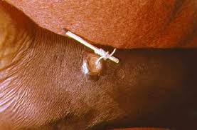
A child with ocularis larva migrans.
If the parasites wander to the eyes, the resulting infection is called ocularis larva migrans.  This tends to happen in older children and young adults.  Symptoms for this form include red eyes, decreased visual clarity, a whitening of the pupil (leukokoria), granulomas, chorioretinitis (inflammation of the choriod and the retina), and even retinal detachment.
Children can also develop a form known as covert toxocariasis, which can result in coughing, abdominal pain, difficulty sleeping, wheezing, or headaches.  This form develops due to chronic exposure.
Because infection is so prevalent in canids in the U.S., human exposure risks are very high here.  Most cases go unrecognized or unreported.  However, about 10,000 cases occur annually.  Somewhere between 4% and 8% of children test positive in serological tests for toxocariasis. Toxocariasis is most prevalent in children ages 1-3, and boys are at a higher risk of infection that girls. (Probably due to an increased probability of fecal-oral contamination since kids this age aren’t exactly well-known for their impeccable hygiene.)

Diagnosis, Treatment, and Prevention of Toxocariasis
            Diagnosis includes identifying clinical symptoms and demonstrating the presence of adult worms in feces or vomit.  Often times, a fecal examination is needed to confirm a diagnosis in dogs.  For humans, ELISA, PCR, and serological tests are most commonly used for diagnostics.
Ivermectin in paste form.
            Dogs are treated using an antihelminthic drug such as piperazine adipate, diethyl carbamazine, pyrantel, benzimidazole compounds, or ivermectin.
Human infections typically resolve themselves, but where treatment is required, it typically includes the administration of antihelminthic drugs such as albendazole or mebendazole.  In severe cases, corticosteroids may be given or surgery may be required.
Infection can be prevented by giving pets regular de-wormers such as fenbendazole, dichlorvos, and piperazine. (Good for your pooch, and reduces your risk of infecting yourself or your dirty children.)  This is especially important in females because dormant larvae may persist in the tissues for years and can be passed on to pups. Outdoor kennels should have floors that are impervious and easy to clean. (In other words, no dirt floors.) Keep food/water containers and bedding clean.  Make sure that the rodent populations are controlled as rodents can be paratenic hosts for the parasites.  Finally, try to keep your dog from eating feces or soil that may have been contaminated with feces.  Infections can also be prevented by owners who take the initiative to clean up after their pets, and by always washing hands thoroughly with soap and water following the handling of dog feces…especially before handling food.  Also, try not to let your children eat dog poop or dirt that could have come into contact with dog poop.

Moral of the Story
            Don’t be the d-bag who is too good to clean up after his/her Toxocara-infested mutt! Keep your pups and kiddos from eating poop and/or dirt and keep your kennels cleaned! (Dog and kid-kennels alike! J/k…Scrubs reference? Anyone?) Also keep Fluffy up to date with de-wormers, and wash your hands if you are playing with his poop. So, watch your children, watch your pets, and wash, wash, wash your hands!

Tuesday, May 22, 2012

Pediculus humanus

LET'S TALK ABOUT LICE! 
Photo of a mother picking the nits off of her child.
     Who doesn't love talking about these creatures? Better yet, who doesn't start obsessively scratching their head when talking about these creatures? (I'm sure that I'll have more people who have raised their hands to the latter question.) Pediculus humanus is found on humans and exists as one of two distinct subspecies: Pediculus humanus capitis (better known as the head louse) and Pediculus humanus humanus (better known as the body louse). This little guy has even gone so far as to invade the way we speak. The term "lousy" derives from the act of one being infested with lice (the plural form of the word louse, if you haven't caught that already). You may also have heard about people being "nit-picky". This comes from the eggs (also known as "nits") that are laid by these animals. The treatment for a lice infestation is to remove the nits, which could be a tedious process before the days of lice combs and treatment shampoos. This is why people who are said to be meticulous are termed "nit-picky".


P. humanus capitis

P. humanus humanus
Taxonomy
      This species of lice is of course an animal belonging in the class Insecta under the phylum Arthropoda. It belongs in the order Phthiraptera with over 3,000 other species of lice. All members of this order are wingless ectoparasites of many different types of birds and mammals. Most lice are scavengers, feeding on dead skin and other debris, but P. humanus is one of the species that takes blood meals. There are four suborders currently recognized under order Phthiraptera: Anoplura (sucking lice found only on mammals), Rhyncothirina (found on elephants and warthogs), Ischnocera (chewing lice that parasitize mostly birds, save for one family that is found on mammals), and Amblycera (more chewing lice that parasitize mostly birds, but are found on some South American and Austrailian mammals). P. humanus belongs to suborder Anoplura. It belongs in family Pediculidae. The two subspecies of P. humanus will interbreed under lab conditions, but they do not interbreed in nature as they occupy very different niches on their hosts.

Copulation in P. humanus humanus (Female on top)

Life Cycle
     The  life cycle for Pediculus humanus involves three distinct stages. The eggs, or "nits", are laid on either the bases of hairs on the head (as in P. humanus capitis) or on the seams of clothing, particularly around the waistline or armpit areas of clothing (as in P. humanus humanus). After a week or two, the nits hatch and release little nymphs. These nymphs look identical to adults except that they lack mature genitals and are smaller in size. The nymphs feed on the host's blood. They nymphs will undergo 3 molts before maturing into adults after 9-12 days. As adults, they will mate and reproduce. As adults, the lice continue to live on blood meals from the host. If they become separated from the host, or if the host dies, the lice will die after a short while living at room temperature.


Pediculosis (Being infected with lice.)
Nits of P. humanus capitis
      A person becomes infected with lice through contact with others who have been infected or through sharing clothing or bedding that is infested. (Pediculosis can also refer to pubic or "crab" lice [Pthirus pubis] infections, but I did not cover that species in this post.) This is particularly problematic in places of high population, but substandard living conditions. This often occurs in transient populations of homeless individuals without access to regular bathing and clean clothing. Historically, it has been problematic for armies or prisoners exposed to substandard living conditions.

       Head lice are not known to carry any diseases, but do cause itchy scalps. Head lice are often found in children and spread quickly in schools and daycares. Head lice are able to live for about two
days off of their host in things like hats, hairbrushes, and pillows.
Nits of P. humanus humanus
     Body lice also cause itching, but unlike head lice, this subspecies is known to serve as a vector for several diseases. This little guy is responsible for louse-borne typhus and louse-borne relapsing fever. Louse-borne typhus has been largely eradicated in many parts of the world, but has existed, and continues to exist in impoverished areas or during times of war or civil unrest.
     The treatment for pediculosis includes an improvement in living conditions, the use of pediculocidal shampoos and lice combs, washing or burning of infected clothing and bedding, and regular bathing.

The Moral of the Story
     Keep yourself clean, wash your cloths, and don't share hats with children or homeless people. :p Being aware of how these animals live is half the battle with preventing or treating their infestations.

Wednesday, May 16, 2012

Dracunculus medinensis

     So, I'm behind again! But here is this week's parasite post! The name of this parasite means "Little Dragon from Medina", which is a throwback to not only its fiery disposition, but also to the high infection rate it once held in the city of Medina. It gets its common name from Carl Linnaeus himself, who first found these creatures in merchants from the West African Coast along the Gulf of Guinea. I first learned about this one years ago in a conversation with a friend about the medical symbol....which is better known as the Rod of Asclepius. This symbol depicts what most people believe is a serpents entwined around a pole. Some people theorize that this is not actually a serpent, but rather a nematode by the common name of "The Guinea Worm". Scientists know this worm as Dracunculus medinensis. This theory is based on the traditional treatment for this worm, which includes slowly extracting the worm by winding it around a stick over a period of days or even weeks. More on this later...let us get to taxonomy!

 Taxonomy
     As mentioned previously, this worm is a member of Phylum Nematoda. It belongs in Class Secernentea along side the Rhabditia, which includes the famous genetic model, C. elegans, and the equally famous Ascaris lumbricoides. D. medinensis is in Order Camallanida, whose members have copepods as their secondary hosts. It is in the Family Dracunculidae and the Genus Dracunculus. There are other members of this genus that infect dogs (D. insignis), otters (D. lutrae), opossums (D. fuelliborni), and even reptiles (D. ophidensis). The most medically important one is, you guessed it, D. medinensis. This bad boy has been found infecting humans, dogs, and even horses and cattle.

D. medinensis Larvae

A copepod (Cyclops) that acts as an
intermediate host for D. medinensis
Life Cycle
       Like all my favorite parasites, this one manipulates the behavior of its host. The guinea worm infects a human host who drinks water that has been contaminated by the worm's intermediate host, a copepod. The copepod dies inside the human's body and releases stage 3 larvae into the stomach. The larvae will move through stomach and intestinal walls to make their way into the abdominal cavity where they mature and mate. The males die after mating, but the females move into the subcutaneous tissues of the body. The female will then form a blister on the skin, usually somewhere on the foot or lower leg. This process takes about a year. The blister will then break open causing severe burning pain. This pain drives the human host to seek water in which to soothe the burning. Once in water, the female is able to sense the change in temperature, and she emerges to release her stage 1 larvae into the liquid environment. The female, which can be as long as 31 inches in length, then suffers the fate of her 1 inch long male companions and dies. The larvae she released into the water are ingested by copepods, in which they molt twice before becoming infectious. 


 Dracunculiasis
     This is also known as "Guinea worm disease" (appropriately) and is only caused by the female of the species. (As you probably could tell from reading the life cycle.) As stated,a blister usually manifests on the lower extremities, but cases have been reported of dracunculiasis on the buttocks, torso, arms, and even on the genitals. (Ouch!) The blister can cause extreme pain and a burning sensation due to the fact that they elicit allergic reactions within the body. This produces rashes, diarrhea, nausea, edema, and dizziness. These reactions subside when the blister ruptures, but then you have to worry about skin ulcers forming and getting the dead worm out. The worms are removed by slowly winding the worms around a stick to extract them over a period of days or weeks. This is because yanking the worm out causes the worm to break, releasing chemicals that have the potential to cause fatalities. The worm can now be removed surgically, but only if the worm is near the skin's surface. Dead worms in joints can cause arthritis and even paralysis if near the spinal cord.


The Moral of the Story
       It goes without saying that you shouldn't drink water of questionable origins, but let's say it anyway. Don't drink unfiltered water from questionable sources. Fine mesh nylon is useful for straining larvae infected copepods and several larvacides have been used to treat water sources that may house these little devils. Many organizations have worked tirelessly to eradicate D. medinensis from the worm's home areas of Africa and Asia. Much progress has been made, but there is still work to be done. We can also learn that sometimes it takes patience to rid yourself of parasites...like spending weeks wrapping a worm from your leg around a stick when all you probably want to do is rip that sucker out an call it a day!


Wednesday, May 9, 2012

Myrmeconema neotropicum

    Since I missed a week, here is my obligatory make-up post.  This parasite has fascinated me since I first learned about it while researching examples of mimicry for an entomology presentation. It has the most amazing life cycle, which involves behavioral modification. W00T! Anyway, I give you...Mymeconema neotropicum!


Taxonomy
     This little nematode (Phylum Nematoda) belongs to class Enoplia alongside whipworms and the ever-awesome Trichinella spiralis. M. neotropicum belongs in the order Mermithida, which has members that are mostly arthropod parasites.  All members of this order have a stichosome.  There are two families within this order. The family that M. neotropicum belongs to is the family Tetradonematidae.

Life Cycle
      This parasite has an interesting life cycle that begins with a frugivorus (fruit-eating) bird ingesting an infected ant of the species Cephalotes atratus. After going through the bird's gut, the parasite eggs are passed out of the birth through defecation. The bird feces are gathered by worker ants of the aforementioned species, and then fed to the ant larvae.  Once inside of the immature ant gut, the parasites hatch from their eggs and migrate to the gaster of the ant, where the parasites mature. Female nematodes are larger than the males in this species.
     About the time that the ant larvae pupate, the now-mature nematodes begin to mate inside of the gaster. The males die shortly after copulating and the females begin to develop eggs within their bodies.  After the ant matures into a young adult, the embryos within the eggs somehow turn the ant gaster translucent, which allows the red-colored embryos to shine through. The longer the ant is infected, the more red the abdomen becomes.
     The parasites also induce a behavioral change. The infected ants carry their abdomens up high in an awkward, unnatural position. The infected ants also move much slower than their healthy brethren. It has been noted that the infected ants begin to forage outside of the nest rather than tending brood within the nest itself as uninfected ants do.
     These behavioral changes coupled with the reddening of the abdomen are an example of what has come to be known as "fruit mimicry", because the ant becomes reminiscent of small red berries.  Our frugivorus bird friends then mistakenly eat the fruit-mimicking antes and the life cycle of the parasite is completed.


Moral of the Story
     This parasite is obviously no threat to us humans, but it is interesting! In fact, I find it more interesting to think of the chain of events that led to this amazing adaptive strategy! THIS is one of the many reasons why parasite evolution is so COOL! :) An now, for your viewing pleasure, a couple of ant pictures. The first is a comparison of an uninfected ant and its infected brother. The second depicts an infected ant alongside some of the berries that are mimicked because of Myrmeconema neotropicum. Enjoy!


Sunday, May 6, 2012

Toxoplasma gondii

      This has been a parasite that I have found interesting since the first time I ever heard of it. I am drawn to this parasite because of its ability to modify the behavior of its host. There is good documentation that this phenomenon occurs in rats, but little is known about how it effects humans.  Some report an increase in thrill-seeking behaviors such as randomly taking up sky-diving or B.A.S.E. jumping post-infection. But I have yet to read any scientific papers that can back up these claims. However, the thought is still pretty interesting!

Taxonomy 
     This parasite is in the phylum apicomplexa, which is in the kingdom chromalveolata. Like other apicomplexans (Plasmodium, Leishmania, Eimeria, etc.), Toxoplasma gondii is parasitic. It is a type of coccidian parasite, belonging to class conoidasida, subclass coccidiasina, and order eucoccidiorida. (Another famous coccidian you may have heard of is Eimeria tenella, the bane of the commercial poultry industry.) T. gondii belongs to the family Sarcocystidae. This parasite was first described in 1908 by Charles Nicolle and Louis Manceaux from a gundi (Ctenodactylus gundi) and by Alfonso Splendore from rabbits that same year.


Life Cycle
    The life cycle of this parasite typically involves two hosts: a cat and a mouse/rat. However, the parasite has been known to opportunistically infect birds and mammals other than cats and mice. It has even been found to infect humans. For the sake of simplicity, I'll describe the life cycle using the cat/mouse model, then discuss human infections.
     To begin, an uninfected feline must ingest an infected mouse. In the infected mouse (the intermediate host), the parasite has formed cysts in the brain, the liver, and the muscles. After ingestion of these cysts, the cyst breaks open to release bradyzoites (non-motile, slow-growing forms) in the stomach. These bradyzoites differentiate to form both asexual tachyzoites (motile, fast-growing forms) and sexual gametocytes (gamete-forming cells). The gametocytes will fuse to form zygotes, which mature into oocysts that will be passed out of the cat with the feces. Instead of eating a cyst, the cat could also ingest oocysts themselves. In this case, the oocysts would release sporozoites that become tachyzoites.
     If the cysts or oocysts are eaten by the intermediate host, in our case the mouse, then bradyzoites and tachyzoites are released in the stomach. Bradyzoites will become tachyzoites in hosts other than cats. The tachyzoites invade cells and multiply via endodyogeny before lysing the cells to release more tachyzoites. During endodyogeny, two daughter cells are created within the mother cell during division. The mother cell is later consumed by the daughter cells just before the daughter cells separate. The tachyzoites are often kept at bay or even destroyed by the host's immune response, but some do manage to undergo transformation into the bradyzoite form. Whenever tachyzoites transform back into bradyzoites, they form cysts in the body tissues of the intermediate host. Because the parasites are within host tissue cells, the host's immune response doesn't kick in to destroy the parasites.


     One of the more interesting aspects of the life cycle is the change in the behavior of the intermediate hosts. Mice and rats have been known to be more bold...by this I mean that they are more likely to run out in front of cats rather than avoiding them. Lab studies have shown that infected mice have less of an aversion to cat urine than do healthy mice. This behavior makes them more likely to be eaten by cats, thus perpetuating the life cycle of the parasite. I do not know whether this parasite somehow suppresses the inhibitions of the mice or if it simply destroys part of the olfactory tissue, but I'd certainly like to know more about the mechanism by which it modifies its host's behaviors.  Humans infected have been said to become more pulled to engage in thrill-seeking behaviors, but I haven't read any scientific studies that support this claim.
     In order for human infection to occur, one must ingest undercooked meat (from say a bird, sheep, or pig) that is tainted with cysts. Another route of infection is the consumption of food or water contaminated by feces from infected cats.  People can also pick up T. gondii via contact with contaminated environments such as fecal-contaminated soils or changing the litter box for an infected cat. Yet another way that people can become infected is via organ transplantation or blood transfusion from infected individuals. Finally, toxoplasmosis can be passed through the placenta from mother to fetus. This is the reason why pregnant women are not supposed to clean out litter boxes.


Toxoplasmosis
     This disease infects approximately 22% of the US population. In non-pregnant individuals it is asymptomatic and often cures itself unless a person becomes immunocompromised. Sometimes it may present with mild flu-like symptoms that persist for several weeks, but eventually goes away. 
     In infected women who become pregnant, the fetus is often protected as the mother has already developed immunities to the parasite. However, if a pregnant woman becomes newly infected, the disease can be congenital (passed to the fetus) through the placental tissue. The severity of the disease varies with the stage of pregnancy, but can result in miscarriage, still birth, or the infant can be born with signs of toxoplasmosis. (Generally, the infant emerges with an abnormally large or abnormally small head.) Sometimes, the infant can be infected at birth and not show signs until later in life. In these cases, infants may develop a loss of vision, a mental disability, or seizures. The loss of vision may be due to a Toxoplasma-induced eye disease in which the retina become irreversibly damaged.
     In immunocompromised people, the disease may experience fevers, headaches, nausea, a decrease in coordination, seizures, and/or confusion. People with HIV are very much at risk for toxoplasmosis.
     This disease is often treated with pyrimethamine and/or sulfadiazine when treatment is needed. AIDS patients with toxoplasmosis may need to continue treatment for the rest of their lives, or for as long as they are immunosuppressed. It is difficult to eradicate, especially in pregnant women and infants due to the nature of the parasites. Many parasites can be killed, but if even a few remain, they are prolific enough to cause a resurgence in their population within the host to the point of pre-treatment levels of infection.

Moral of the Story
     The best way to prevent yourself from getting toxoplasmosis is to reduce the risks from food and environmental contamination. Always cook your meat thoroughly and allow adequate resting time prior to carving. (You can also freeze the meat prior to cooking to further reduce the risk of infection.) Always wash and/or peel fruits and vegetables before consumption. Don't drink water from questionable sources. Be sure to wash cutting boards and utensils thoroughly. Wear gloves while gardening and keep  sandboxes for children covered when not in use. Feed your kitty food that you know isn't contaminated and keep them indoors to reduce their chances of picking up toxoplasmosis. And finally, if you are pregnant or immunocompromised, have someone else change out the litter box and avoid handling stray or unfamiliar cats.

Saturday, April 21, 2012

Leishmania mexicana

     Today, I helped a friend attempt to catch woodrats (Neotoma micropus) and sandflies. She is working on her thesis....she's on the hunt for a particular parasite.  This parasite isn't a big problem in this area, but it is a problem in other areas. She's looking to see if it is here...and for good reason. In 2007, dermatologists in Dallas, Texas diagnosed NINE cases of cutaneous leishmaniasis...but we will get to that later. First things first, taxonomy!

Taxonomy
L. mexicana Promastigote
     This parasite is a protist withing the excavate clade. Leishmania in the phylum Euglenozoa....which contains the commonly known free-living Euglena; an elegant example of a flagellate often found in pond scum.  Leishmania belongs in class Kinetoplastida and order Trypanosomatida alongside other trypanosomes such as Trypanosoma cruzi (which causes Chagas' disease) and Trypanosoma gambiense (which causes sleeping sickness).  The genus, Leishmania, has many pathogenic species. The Leishmania donovani species complex causes the most terrible form of this disease, which is known as visceral leishmaniasis (a.k.a. "Kala-Azar"). Leishmania braziliensis causes mucocutaneous leishmaniasis. Many other species cause cutaneous leishmaniasis, but only one is found in the new world....this is Leishmania mexicana.

L. mexicana Amastigote (in green)

Life Cycle
     The life cycle of this organism involves two body forms. An infected female sandfly takes a blood meal from an uninfected human. During this feeding, Leishmania mexicana promastigotes make their way into their new host's blood stream. As the body's immune system goes to work, macrophages ingest the promastigotes. While inside of the macrophages, the promastigotes transform into an amastigote form (the green ovoid organism in the picture on the right). The amastigotes reproduce within the macrophages and other cells. Then, along comes an innocent, uninfected female sandfly hungry for a delicious blood meal. As the sandfly sucks the blood of the infected human, she ingests the infected macrophages. Once inside of the sandfly, the amastigotes burst from the macrophages and transform into the promastigote stage within the midgut of the sandfly. Then the promastigotes migrate to the proboscis of the fly, where they await for the fly to have its next blood meal...completing the life cycle.

Cutaneous and Diffused Cutaneous Leishmaniasis
     These forms of leishmaniasis are less dangerous than visceral forms. The cutaneous form manifests as ulcers at the site where the sandfly bites its host. In this form, the amastigotes don't spread to other areas. The ulcers show up anywhere from a few days to a few months post-infection. This form usually heals on its own given time. 

     The diffused form of cutaneous leishmaniasis only manifests after the amastigotes cutaneously spread due to defective immune cells (such as T-cells). This can cause sores and ulcers all over the body that do not respond well to drugs. Penstostam is a drug that is often used in the US to treat this form of leishmaniasis. Penstostam works by inhibiting ATP synthesis.  There is ongoing research into vaccine development that works against the promastigote life stage.
     These diseases are typically only found in Central and South America. However, there have been cases reported right here in Texas. Cases began appearing in South Texas years ago, but it wasn't until the last few years that it was reported further north. In 2007, nine different cases were reported from around the Dallas area. Researchers believe that sandflies infect woodrats as they search for blood meals. These woodrats then become what we call a "reservoir". This means that they are able to serve as a sort of storage space for the propagation of the Leishmania mexicana protozoans without a human host. This keeps the protists circulating in the sandflies, making them potential disease vectors.

Moral of the Story
     There is strong evidence to suggest that if sandflies and woodrats are present, so could be present this parasite. People in rural areas are more at risk despite the sandflies' recent movements into more urban areas. Using insect repellants will reduce the risk of being bitten, so keep that in mind the next time you head out to the boonies! :p

Saturday, April 14, 2012

Naegleria fowleri

So for my first post, I thought it would be appropriate to start with something small. One of my friends studied this organism for her thesis work and her defense is next week...so it seemed like a great place to begin!

Taxonomy
      This protozoan is a member of phylum Percolozoa because they are able to transform between amoeboid, flagellate, and encysted stages. These organisms also belong to class heterolobosea, which is closely related to the euglenozoans.  They belong to order schizopyrenida and family vahlkampfidae. The genus Naegleria was named after Mathiew Naegler, a zoologist from France.

Life Cycle
      The cysts of N. fowleri form from the trophozoite stage under harsh conditions, such as overpopulation, limited availability of food, cold temperatures (below 10°C), desiccation, etc.When temperatures rise (especially after they reach 42°C), the free-living, reproductive life stage (the trophozoite) undergoes binary fission. The trophozoite moves via pseduopodia and feeds on bacteria within freshwater lakes, rivers, or hot springs. When these trophozoites become exposed to changes in ionic concentrations of their environment, they transform into the biflagellate body form within minutes of said exposure.


     This organism may opportunistically infect humans by entering the nose of unwary swimmers while existing in the flagellate stage.  Most often, significant increases in temperature and/or a reduced amount of rainfall can induce pre-existing trophozoites to enter into the flagellate form. After entering a human host, this amoeba travels to the brain and proceeds to inflame the meninges.


Primary Amoebic Meningoencephalitis

      PAM was first documented in 1965 from Austrailia.  The causative agent was not identified until 1968. Between 1963 and 1965, 16 cases were reported from Usti nad Labem in the Czech Republic. The amoeba finally was given the name Naegleria fowleri in 1970. Recent cases include a 7-year-old girl from Stillwater, Minnesota in 2010 and a 16-year-old-girl from Mims, Florida in August 2011. Two individuals from Louisiana contracted the parasite in December 2011 after inhaling infected tap water.  These individuals were using a neti pot.      
      This disease affects the central nervous system. It begins with N. fowleri entering the nasal cavity of a human host and attaching itself to the olfactory nerve. It then makes its way into the olfactory bulbs within the forebrain and begins to feed on nerve tissue and to multiply. 3-7 days post infection, patients begin having trouble properly identifying smells (a symptom called "parosmia") and eventually lose the ability to detect odors at all ("anosmia"). This will often lead to an inability to detect the basic taste sensations (known as having "ageusia"). As the olfactory bulb nerve cells are eaten, necrotic lesions begin to develop. 
      The infection quickly progresses to the rest of the cerebrum, which causes an inflammation of the meninges. During this time, patients experience headaches (a.k.a. "cephalgia"), stiff neck muscles, nausea, vomiting, seizures, delirium, and eventually coma. Once the infection spreads to the brain stem and begins feasting on the cells of the medulla oblongata (about 14 days post-infection), death usually results from respiratory failure.
      Infection by this protist is extremely rare and this disease has a mortality rate of approximately 97%. Due to the rapid progression of the disease and the symptoms that are easily mistaken for symptoms of other problems, positive diagnosis is difficult. To diagnose, microbial culturing from CSF is needed, but if PAM is suspect, treatment will often begin prior to confirmed diagnosis. The battery of antiamoebic drugs that are typically administered are somewhat dangerous taken together and this is often not a pleasant experience for the patient.

Moral of the Story
       Don't swim in stagnant water, especially during droughts or particularly hot summers! And always heed warnings...you are NOT immune to Naegleria infection!