Pages

Tuesday, May 22, 2012

Pediculus humanus

LET'S TALK ABOUT LICE! 
Photo of a mother picking the nits off of her child.
     Who doesn't love talking about these creatures? Better yet, who doesn't start obsessively scratching their head when talking about these creatures? (I'm sure that I'll have more people who have raised their hands to the latter question.) Pediculus humanus is found on humans and exists as one of two distinct subspecies: Pediculus humanus capitis (better known as the head louse) and Pediculus humanus humanus (better known as the body louse). This little guy has even gone so far as to invade the way we speak. The term "lousy" derives from the act of one being infested with lice (the plural form of the word louse, if you haven't caught that already). You may also have heard about people being "nit-picky". This comes from the eggs (also known as "nits") that are laid by these animals. The treatment for a lice infestation is to remove the nits, which could be a tedious process before the days of lice combs and treatment shampoos. This is why people who are said to be meticulous are termed "nit-picky".


P. humanus capitis

P. humanus humanus
Taxonomy
      This species of lice is of course an animal belonging in the class Insecta under the phylum Arthropoda. It belongs in the order Phthiraptera with over 3,000 other species of lice. All members of this order are wingless ectoparasites of many different types of birds and mammals. Most lice are scavengers, feeding on dead skin and other debris, but P. humanus is one of the species that takes blood meals. There are four suborders currently recognized under order Phthiraptera: Anoplura (sucking lice found only on mammals), Rhyncothirina (found on elephants and warthogs), Ischnocera (chewing lice that parasitize mostly birds, save for one family that is found on mammals), and Amblycera (more chewing lice that parasitize mostly birds, but are found on some South American and Austrailian mammals). P. humanus belongs to suborder Anoplura. It belongs in family Pediculidae. The two subspecies of P. humanus will interbreed under lab conditions, but they do not interbreed in nature as they occupy very different niches on their hosts.

Copulation in P. humanus humanus (Female on top)

Life Cycle
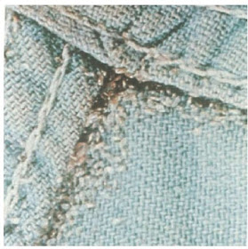
     The  life cycle for Pediculus humanus involves three distinct stages. The eggs, or "nits", are laid on either the bases of hairs on the head (as in P. humanus capitis) or on the seams of clothing, particularly around the waistline or armpit areas of clothing (as in P. humanus humanus). After a week or two, the nits hatch and release little nymphs. These nymphs look identical to adults except that they lack mature genitals and are smaller in size. The nymphs feed on the host's blood. They nymphs will undergo 3 molts before maturing into adults after 9-12 days. As adults, they will mate and reproduce. As adults, the lice continue to live on blood meals from the host. If they become separated from the host, or if the host dies, the lice will die after a short while living at room temperature.


Pediculosis (Being infected with lice.)
Nits of P. humanus capitis
      A person becomes infected with lice through contact with others who have been infected or through sharing clothing or bedding that is infested. (Pediculosis can also refer to pubic or "crab" lice [Pthirus pubis] infections, but I did not cover that species in this post.) This is particularly problematic in places of high population, but substandard living conditions. This often occurs in transient populations of homeless individuals without access to regular bathing and clean clothing. Historically, it has been problematic for armies or prisoners exposed to substandard living conditions.

       Head lice are not known to carry any diseases, but do cause itchy scalps. Head lice are often found in children and spread quickly in schools and daycares. Head lice are able to live for about two
days off of their host in things like hats, hairbrushes, and pillows.
Nits of P. humanus humanus
     Body lice also cause itching, but unlike head lice, this subspecies is known to serve as a vector for several diseases. This little guy is responsible for louse-borne typhus and louse-borne relapsing fever. Louse-borne typhus has been largely eradicated in many parts of the world, but has existed, and continues to exist in impoverished areas or during times of war or civil unrest.
     The treatment for pediculosis includes an improvement in living conditions, the use of pediculocidal shampoos and lice combs, washing or burning of infected clothing and bedding, and regular bathing.

The Moral of the Story
     Keep yourself clean, wash your cloths, and don't share hats with children or homeless people. :p Being aware of how these animals live is half the battle with preventing or treating their infestations.

Wednesday, May 16, 2012

Dracunculus medinensis

     So, I'm behind again! But here is this week's parasite post! The name of this parasite means "Little Dragon from Medina", which is a throwback to not only its fiery disposition, but also to the high infection rate it once held in the city of Medina. It gets its common name from Carl Linnaeus himself, who first found these creatures in merchants from the West African Coast along the Gulf of Guinea. I first learned about this one years ago in a conversation with a friend about the medical symbol....which is better known as the Rod of Asclepius. This symbol depicts what most people believe is a serpents entwined around a pole. Some people theorize that this is not actually a serpent, but rather a nematode by the common name of "The Guinea Worm". Scientists know this worm as Dracunculus medinensis. This theory is based on the traditional treatment for this worm, which includes slowly extracting the worm by winding it around a stick over a period of days or even weeks. More on this later...let us get to taxonomy!

 Taxonomy
     As mentioned previously, this worm is a member of Phylum Nematoda. It belongs in Class Secernentea along side the Rhabditia, which includes the famous genetic model, C. elegans, and the equally famous Ascaris lumbricoides. D. medinensis is in Order Camallanida, whose members have copepods as their secondary hosts. It is in the Family Dracunculidae and the Genus Dracunculus. There are other members of this genus that infect dogs (D. insignis), otters (D. lutrae), opossums (D. fuelliborni), and even reptiles (D. ophidensis). The most medically important one is, you guessed it, D. medinensis. This bad boy has been found infecting humans, dogs, and even horses and cattle.

D. medinensis Larvae

A copepod (Cyclops) that acts as an
intermediate host for D. medinensis
Life Cycle
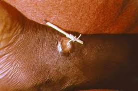
       Like all my favorite parasites, this one manipulates the behavior of its host. The guinea worm infects a human host who drinks water that has been contaminated by the worm's intermediate host, a copepod. The copepod dies inside the human's body and releases stage 3 larvae into the stomach. The larvae will move through stomach and intestinal walls to make their way into the abdominal cavity where they mature and mate. The males die after mating, but the females move into the subcutaneous tissues of the body. The female will then form a blister on the skin, usually somewhere on the foot or lower leg. This process takes about a year. The blister will then break open causing severe burning pain. This pain drives the human host to seek water in which to soothe the burning. Once in water, the female is able to sense the change in temperature, and she emerges to release her stage 1 larvae into the liquid environment. The female, which can be as long as 31 inches in length, then suffers the fate of her 1 inch long male companions and dies. The larvae she released into the water are ingested by copepods, in which they molt twice before becoming infectious. 


 Dracunculiasis
     This is also known as "Guinea worm disease" (appropriately) and is only caused by the female of the species. (As you probably could tell from reading the life cycle.) As stated,a blister usually manifests on the lower extremities, but cases have been reported of dracunculiasis on the buttocks, torso, arms, and even on the genitals. (Ouch!) The blister can cause extreme pain and a burning sensation due to the fact that they elicit allergic reactions within the body. This produces rashes, diarrhea, nausea, edema, and dizziness. These reactions subside when the blister ruptures, but then you have to worry about skin ulcers forming and getting the dead worm out. The worms are removed by slowly winding the worms around a stick to extract them over a period of days or weeks. This is because yanking the worm out causes the worm to break, releasing chemicals that have the potential to cause fatalities. The worm can now be removed surgically, but only if the worm is near the skin's surface. Dead worms in joints can cause arthritis and even paralysis if near the spinal cord.


The Moral of the Story
       It goes without saying that you shouldn't drink water of questionable origins, but let's say it anyway. Don't drink unfiltered water from questionable sources. Fine mesh nylon is useful for straining larvae infected copepods and several larvacides have been used to treat water sources that may house these little devils. Many organizations have worked tirelessly to eradicate D. medinensis from the worm's home areas of Africa and Asia. Much progress has been made, but there is still work to be done. We can also learn that sometimes it takes patience to rid yourself of parasites...like spending weeks wrapping a worm from your leg around a stick when all you probably want to do is rip that sucker out an call it a day!


Wednesday, May 9, 2012

Myrmeconema neotropicum

    Since I missed a week, here is my obligatory make-up post.  This parasite has fascinated me since I first learned about it while researching examples of mimicry for an entomology presentation. It has the most amazing life cycle, which involves behavioral modification. W00T! Anyway, I give you...Mymeconema neotropicum!


Taxonomy
     This little nematode (Phylum Nematoda) belongs to class Enoplia alongside whipworms and the ever-awesome Trichinella spiralis. M. neotropicum belongs in the order Mermithida, which has members that are mostly arthropod parasites.  All members of this order have a stichosome.  There are two families within this order. The family that M. neotropicum belongs to is the family Tetradonematidae.

Life Cycle
      This parasite has an interesting life cycle that begins with a frugivorus (fruit-eating) bird ingesting an infected ant of the species Cephalotes atratus. After going through the bird's gut, the parasite eggs are passed out of the birth through defecation. The bird feces are gathered by worker ants of the aforementioned species, and then fed to the ant larvae.  Once inside of the immature ant gut, the parasites hatch from their eggs and migrate to the gaster of the ant, where the parasites mature. Female nematodes are larger than the males in this species.
     About the time that the ant larvae pupate, the now-mature nematodes begin to mate inside of the gaster. The males die shortly after copulating and the females begin to develop eggs within their bodies.  After the ant matures into a young adult, the embryos within the eggs somehow turn the ant gaster translucent, which allows the red-colored embryos to shine through. The longer the ant is infected, the more red the abdomen becomes.
     The parasites also induce a behavioral change. The infected ants carry their abdomens up high in an awkward, unnatural position. The infected ants also move much slower than their healthy brethren. It has been noted that the infected ants begin to forage outside of the nest rather than tending brood within the nest itself as uninfected ants do.
     These behavioral changes coupled with the reddening of the abdomen are an example of what has come to be known as "fruit mimicry", because the ant becomes reminiscent of small red berries.  Our frugivorus bird friends then mistakenly eat the fruit-mimicking antes and the life cycle of the parasite is completed.


Moral of the Story
     This parasite is obviously no threat to us humans, but it is interesting! In fact, I find it more interesting to think of the chain of events that led to this amazing adaptive strategy! THIS is one of the many reasons why parasite evolution is so COOL! :) An now, for your viewing pleasure, a couple of ant pictures. The first is a comparison of an uninfected ant and its infected brother. The second depicts an infected ant alongside some of the berries that are mimicked because of Myrmeconema neotropicum. Enjoy!


Sunday, May 6, 2012

Toxoplasma gondii

      This has been a parasite that I have found interesting since the first time I ever heard of it. I am drawn to this parasite because of its ability to modify the behavior of its host. There is good documentation that this phenomenon occurs in rats, but little is known about how it effects humans.  Some report an increase in thrill-seeking behaviors such as randomly taking up sky-diving or B.A.S.E. jumping post-infection. But I have yet to read any scientific papers that can back up these claims. However, the thought is still pretty interesting!

Taxonomy 
     This parasite is in the phylum apicomplexa, which is in the kingdom chromalveolata. Like other apicomplexans (Plasmodium, Leishmania, Eimeria, etc.), Toxoplasma gondii is parasitic. It is a type of coccidian parasite, belonging to class conoidasida, subclass coccidiasina, and order eucoccidiorida. (Another famous coccidian you may have heard of is Eimeria tenella, the bane of the commercial poultry industry.) T. gondii belongs to the family Sarcocystidae. This parasite was first described in 1908 by Charles Nicolle and Louis Manceaux from a gundi (Ctenodactylus gundi) and by Alfonso Splendore from rabbits that same year.


Life Cycle
    The life cycle of this parasite typically involves two hosts: a cat and a mouse/rat. However, the parasite has been known to opportunistically infect birds and mammals other than cats and mice. It has even been found to infect humans. For the sake of simplicity, I'll describe the life cycle using the cat/mouse model, then discuss human infections.
     To begin, an uninfected feline must ingest an infected mouse. In the infected mouse (the intermediate host), the parasite has formed cysts in the brain, the liver, and the muscles. After ingestion of these cysts, the cyst breaks open to release bradyzoites (non-motile, slow-growing forms) in the stomach. These bradyzoites differentiate to form both asexual tachyzoites (motile, fast-growing forms) and sexual gametocytes (gamete-forming cells). The gametocytes will fuse to form zygotes, which mature into oocysts that will be passed out of the cat with the feces. Instead of eating a cyst, the cat could also ingest oocysts themselves. In this case, the oocysts would release sporozoites that become tachyzoites.
     If the cysts or oocysts are eaten by the intermediate host, in our case the mouse, then bradyzoites and tachyzoites are released in the stomach. Bradyzoites will become tachyzoites in hosts other than cats. The tachyzoites invade cells and multiply via endodyogeny before lysing the cells to release more tachyzoites. During endodyogeny, two daughter cells are created within the mother cell during division. The mother cell is later consumed by the daughter cells just before the daughter cells separate. The tachyzoites are often kept at bay or even destroyed by the host's immune response, but some do manage to undergo transformation into the bradyzoite form. Whenever tachyzoites transform back into bradyzoites, they form cysts in the body tissues of the intermediate host. Because the parasites are within host tissue cells, the host's immune response doesn't kick in to destroy the parasites.


     One of the more interesting aspects of the life cycle is the change in the behavior of the intermediate hosts. Mice and rats have been known to be more bold...by this I mean that they are more likely to run out in front of cats rather than avoiding them. Lab studies have shown that infected mice have less of an aversion to cat urine than do healthy mice. This behavior makes them more likely to be eaten by cats, thus perpetuating the life cycle of the parasite. I do not know whether this parasite somehow suppresses the inhibitions of the mice or if it simply destroys part of the olfactory tissue, but I'd certainly like to know more about the mechanism by which it modifies its host's behaviors.  Humans infected have been said to become more pulled to engage in thrill-seeking behaviors, but I haven't read any scientific studies that support this claim.
     In order for human infection to occur, one must ingest undercooked meat (from say a bird, sheep, or pig) that is tainted with cysts. Another route of infection is the consumption of food or water contaminated by feces from infected cats.  People can also pick up T. gondii via contact with contaminated environments such as fecal-contaminated soils or changing the litter box for an infected cat. Yet another way that people can become infected is via organ transplantation or blood transfusion from infected individuals. Finally, toxoplasmosis can be passed through the placenta from mother to fetus. This is the reason why pregnant women are not supposed to clean out litter boxes.


Toxoplasmosis
     This disease infects approximately 22% of the US population. In non-pregnant individuals it is asymptomatic and often cures itself unless a person becomes immunocompromised. Sometimes it may present with mild flu-like symptoms that persist for several weeks, but eventually goes away. 
     In infected women who become pregnant, the fetus is often protected as the mother has already developed immunities to the parasite. However, if a pregnant woman becomes newly infected, the disease can be congenital (passed to the fetus) through the placental tissue. The severity of the disease varies with the stage of pregnancy, but can result in miscarriage, still birth, or the infant can be born with signs of toxoplasmosis. (Generally, the infant emerges with an abnormally large or abnormally small head.) Sometimes, the infant can be infected at birth and not show signs until later in life. In these cases, infants may develop a loss of vision, a mental disability, or seizures. The loss of vision may be due to a Toxoplasma-induced eye disease in which the retina become irreversibly damaged.
     In immunocompromised people, the disease may experience fevers, headaches, nausea, a decrease in coordination, seizures, and/or confusion. People with HIV are very much at risk for toxoplasmosis.
     This disease is often treated with pyrimethamine and/or sulfadiazine when treatment is needed. AIDS patients with toxoplasmosis may need to continue treatment for the rest of their lives, or for as long as they are immunosuppressed. It is difficult to eradicate, especially in pregnant women and infants due to the nature of the parasites. Many parasites can be killed, but if even a few remain, they are prolific enough to cause a resurgence in their population within the host to the point of pre-treatment levels of infection.

Moral of the Story
     The best way to prevent yourself from getting toxoplasmosis is to reduce the risks from food and environmental contamination. Always cook your meat thoroughly and allow adequate resting time prior to carving. (You can also freeze the meat prior to cooking to further reduce the risk of infection.) Always wash and/or peel fruits and vegetables before consumption. Don't drink water from questionable sources. Be sure to wash cutting boards and utensils thoroughly. Wear gloves while gardening and keep  sandboxes for children covered when not in use. Feed your kitty food that you know isn't contaminated and keep them indoors to reduce their chances of picking up toxoplasmosis. And finally, if you are pregnant or immunocompromised, have someone else change out the litter box and avoid handling stray or unfamiliar cats.